


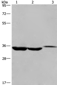
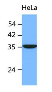
Figure 1. Western blot analysis of ING2 using anti-ING2 antibody (A04290-3). Electrophoresis was performed on a 5-20% SDS-PAGE gel at 70V (Stacking gel) / 90V (Resolving gel) for 2-3 hours. The sample well of each lane was loaded with 30 ug of sample under reducing conditions. Lane 1: human 293T whole cell lysates, Lane 2: human HepG2 whole cell lysates, Lane 3: rat thymus tissue lysates, Lane 4: rat PC-12 whole cell lysates, Lane 5: mouse thymus tissue lysates, Lane 6: mouse RAW264.7 whole cell lysates, Lane 7: mouse EL-4 whole cell lysates, Lane 8: mouse Ana-1 whole cell lysates. After electrophoresis, proteins were transferred to a nitrocellulose membrane at 150 mA for 50-90 minutes. Blocked the membrane with 5% non-fat milk/TBS for 1.5 hour at RT. The membrane was incubated with rabbit anti-ING2 antigen affinity purified polyclonal antibody (Catalog # A04290-3) at 0.5 microg/mL overnight at 4°C, then washed with TBS-0.1%Tween 3 times with 5 minutes each and probed with a goat anti-rabbit IgG-HRP secondary antibody at a dilution of 1:5000 for 1.5 hour at RT. The signal is developed using an Enhanced Chemiluminescent detection (ECL) kit (Catalog # EK1002) with Tanon 5200 system. A specific band was detected for ING2 at approximately 37 kDa. The expected band size for ING2 is at 33 kDa.
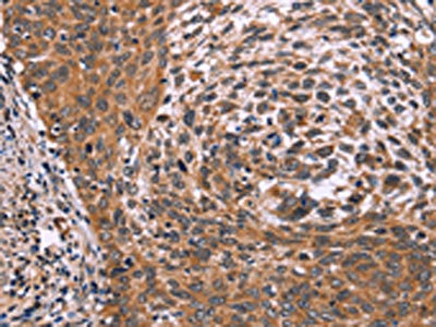
 and anti-Beta Tubulin antibody (M01857-3). ING2 was detected in immunocytochemical section of HELA cell. Enzyme antigen retrieval was performed using IHC enzyme antigen retrieval reagent (AR0022) for 15 mins. The cells were blocked with 10% goat serum. And then incubated with 5 microg/mL rabbit anti-ING2 Antibody (A04290-3) and mouse anti-Beta Tubulin antibody (M01857-3) overnight at 4°C. DyLight®488 Conjugated Goat Anti-Rabbit IgG (BA1127) and Cy3 Conjugated Goat Anti-Mouse IgG (BA1031) were used as secondary antibody at 1:500 dilution and incubated for 30 minutes at 37°C. Visualize using a fluorescence microscope and filter sets appropriate for the label used.")
. Overlay histogram showing HepG2 cells stained with A04290-3 (Blue line). To facilitate intracellular staining, cells were fixed with 4% paraformaldehyde and permeabilized with permeabilization buffer. The cells were blocked with 10% normal goat serum. And then incubated with rabbit anti-ING2 Antibody (A04290-3, 1 microg/1x106 cells) for 30 min at 20°C. DyLight®488 conjugated goat anti-rabbit IgG (BA1127, 5-10 microg/1x106 cells) was used as secondary antibody for 30 minutes at 20°C. Isotype control antibody (Green line) was rabbit IgG (1 microg/1x106) used under the same conditions. Unlabelled sample (Red line) was also used as a control.")
Figure 1. Western blot analysis of ING2 using anti-ING2 antibody (A04290-3). Electrophoresis was performed on a 5-20% SDS-PAGE gel at 70V (Stacking gel) / 90V (Resolving gel) for 2-3 hours. The sample well of each lane was loaded with 30 ug of sample under reducing conditions. Lane 1: human 293T whole cell lysates, Lane 2: human HepG2 whole cell lysates, Lane 3: rat thymus tissue lysates, Lane 4: rat PC-12 whole cell lysates, Lane 5: mouse thymus tissue lysates, Lane 6: mouse RAW264.7 whole cell lysates, Lane 7: mouse EL-4 whole cell lysates, Lane 8: mouse Ana-1 whole cell lysates. After electrophoresis, proteins were transferred to a nitrocellulose membrane at 150 mA for 50-90 minutes. Blocked the membrane with 5% non-fat milk/TBS for 1.5 hour at RT. The membrane was incubated with rabbit anti-ING2 antigen affinity purified polyclonal antibody (Catalog # A04290-3) at 0.5 microg/mL overnight at 4°C, then washed with TBS-0.1%Tween 3 times with 5 minutes each and probed with a goat anti-rabbit IgG-HRP secondary antibody at a dilution of 1:5000 for 1.5 hour at RT. The signal is developed using an Enhanced Chemiluminescent detection (ECL) kit (Catalog # EK1002) with Tanon 5200 system. A specific band was detected for ING2 at approximately 37 kDa. The expected band size for ING2 is at 33 kDa.
Anti-ING2 Antibody Picoband(r)
A04290-3-CARRIER-FREE
ApplicationsFlow Cytometry, ImmunoFluorescence, Western Blot, ELISA, ImmunoCytoChemistry
Product group Antibodies
ReactivityHuman, Mouse, Rat
TargetING2
Overview
- SupplierBoster Bio
- Product NameAnti-ING2 Antibody Picoband(r)
- Delivery Days Customer9
- ApplicationsFlow Cytometry, ImmunoFluorescence, Western Blot, ELISA, ImmunoCytoChemistry
- CertificationResearch Use Only
- ClonalityPolyclonal
- Concentration500 ug/ml
- Gene ID3622
- Target nameING2
- Target descriptioninhibitor of growth family member 2
- Target synonymsING1L, p33ING2, inhibitor of growth protein 2, ING1Lp, inhibitor of growth 1-like protein, p32
- HostRabbit
- IsotypeIgG
- Protein IDQ9H160
- Protein NameInhibitor of growth protein 2
- Scientific DescriptionBoster Bio Anti-ING2 Antibody Picoband® catalog # A04290-3. Tested in ELISA, IF, ICC, WB, Flow Cytometry applications. This antibody reacts with Human, Mouse, Rat. The brand Picoband indicates this is a premium antibody that guarantees superior quality, high affinity, and strong signals with minimal background in Western blot applications. Only our best-performing antibodies are designated as Picoband, ensuring unmatched performance.
- ReactivityHuman, Mouse, Rat
- Storage Instruction-20°C,2°C to 8°C
- UNSPSC12352203
Related products
Product group Antibodies
Anti-ING2 AntibodyA38672
ApplicationsWestern Blot, ImmunoHistoChemistry
ReactivityHuman, Mouse
- SizePrice
Product group Antibodies

Goat anti-ING2 / ING1LEB05452
ApplicationsWestern Blot, ELISA
ReactivityBovine, Human
TargetING2
- SizePrice
Product group Antibodies
ING2 AntibodyCSB-PA266125
ApplicationsELISA, ImmunoHistoChemistry
ReactivityHuman, Mouse
TargetING2
- SizePrice
Product group Antibodies
ING2 Polyclonal AntibodyBS-16653R
ApplicationsWestern Blot
ReactivityCanine, Equine, Human, Mouse, Porcine, Rabbit, Rat, Sheep
TargetING2
- SizePrice
Product group Antibodies

ING2 AntibodyLS-C400819
ApplicationsELISA, ImmunoHistoChemistry
ReactivityHuman, Mouse
TargetING2
- SizePrice
Product group Antibodies
ING2 antibody [AT39E5]GTX53732
ApplicationsWestern Blot, ELISA
ReactivityHuman
TargetING2
- SizePrice

Product group Antibodies
Anti-ING2 AntibodyHPA019486
ApplicationsImmunoCytoChemistry, ImmunoHistoChemistry
ReactivityHuman
TargetING2
- SizePrice
Product group Antibodies
Anti-ING2Y158058
ApplicationsELISA, ImmunoHistoChemistry
ReactivityHuman, Mouse, Rat
- SizePrice