


Immunohistochemical staining of human thyroid gland shows distinct cytoplasmic and nuclear positivity in glandular cells.
Immunohistochemical staining of human thyroid gland shows distinct cytoplasmic and nuclear positivity in glandular cells.
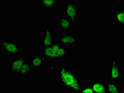
Anti-ING5 Antibody
HPA042685
ApplicationsImmunoHistoChemistry
Product group Antibodies
ReactivityHuman
TargetING5
Overview
- SupplierAtlas Antibodies
- Product NameAnti-ING5 Antibody
- Delivery Days Customer4
- ApplicationsImmunoHistoChemistry
- CertificationResearch Use Only
- ClonalityPolyclonal
- ConjugateUnconjugated
- Gene ID84289
- Target nameING5
- Target descriptioninhibitor of growth family member 5
- Target synonymsp28ING5, inhibitor of growth protein 5
- HostRabbit
- IsotypeIgG
- Protein IDQ8WYH8
- Protein NameInhibitor of growth protein 5
- Scientific DescriptionRecombinant Protein Epitope Signature Tag (PrEST) antigen sequence
- ReactivityHuman
- Storage Instruction-20°C,2°C to 8°C
- UNSPSC41116161
Datasheet
MSDS
Related products
Product group Antibodies
Anti-ING5 Antibody144-07288
ApplicationsImmunoFluorescence, Western Blot
ReactivityHuman
TargetING5
- SizePrice

Product group Antibodies
Anti-ING5 Antibody Picoband(r)A04974-3-CARRIER-FREE
ApplicationsFlow Cytometry, ImmunoFluorescence, Western Blot, ELISA, ImmunoCytoChemistry
ReactivityHuman, Monkey
TargetING5
- SizePrice
Product group Antibodies
Anti-ING5 AntibodyA31978
ApplicationsImmunoFluorescence, Western Blot, ImmunoHistoChemistry
ReactivityHuman, Mouse
- SizePrice
Product group Antibodies

ING5 AntibodyLS-C346332
ApplicationsImmunoFluorescence, Western Blot, ImmunoHistoChemistry
ReactivityHuman
TargetING5
- SizePrice
Product group Antibodies
ING5 AntibodyCSB-PA820185LA01HU
ApplicationsImmunoFluorescence, ELISA
ReactivityHuman
TargetING5
- SizePrice
![ING5 antibody [N2C1], Internal detects ING5 protein by Western blot analysis. A. 30 μg Jurkat whole cell lysate/extract 12 % SDS-PAGE ING5 antibody [N2C1], Internal (GTX102482) dilution: 1:1000](https://www.genetex.com/upload/website/prouct_img/normal/GTX102482/GTX102482_39617_WB_w_23060100_570.webp)
Product group Antibodies
ING5 antibody [N2C1], InternalGTX102482
ApplicationsWestern Blot
ReactivityHuman
TargetING5
- SizePrice