


Figure 1. Western blot analysis of Inhibin beta A/INHBA using anti-Inhibin beta A/INHBA antibody (M06777). Electrophoresis was performed on a 5-20% SDS-PAGE gel at 70V (Stacking gel) / 90V (Resolving gel) for 2-3 hours. The sample well of each lane was loaded with 30 ug of sample under reducing conditions. Lane 1: human U2OS whole cell lysates, Lane 2: human PC-3 whole cell lysates, Lane 3: human Hacat whole cell lysates, Lane 4: human U251 whole cell lysates. After electrophoresis, proteins were transferred to a nitrocellulose membrane at 150 mA for 50-90 minutes. Blocked the membrane with 5% non-fat milk/TBS for 1.5 hour at RT. The membrane was incubated with rabbit anti-Inhibin beta A/INHBA antigen affinity purified monoclonal antibody (M06777) at 1:500 overnight at 4°C, then washed with TBS-0.1%Tween 3 times with 5 minutes each and probed with a goat anti-rabbit IgG-HRP secondary antibody at a dilution of 1:500 for 1.5 hour at RT. The signal is developed using an Enhanced Chemiluminescent detection (ECL) kit (Catalog # EK1002) with Tanon 5200 system. A specific band was detected for Inhibin beta A/INHBA at approximately 43 kDa. The expected band size for Inhibin beta A/INHBA is at 47 kDa.
Figure 1. Western blot analysis of Inhibin beta A/INHBA using anti-Inhibin beta A/INHBA antibody (M06777). Electrophoresis was performed on a 5-20% SDS-PAGE gel at 70V (Stacking gel) / 90V (Resolving gel) for 2-3 hours. The sample well of each lane was loaded with 30 ug of sample under reducing conditions. Lane 1: human U2OS whole cell lysates, Lane 2: human PC-3 whole cell lysates, Lane 3: human Hacat whole cell lysates, Lane 4: human U251 whole cell lysates. After electrophoresis, proteins were transferred to a nitrocellulose membrane at 150 mA for 50-90 minutes. Blocked the membrane with 5% non-fat milk/TBS for 1.5 hour at RT. The membrane was incubated with rabbit anti-Inhibin beta A/INHBA antigen affinity purified monoclonal antibody (M06777) at 1:500 overnight at 4°C, then washed with TBS-0.1%Tween 3 times with 5 minutes each and probed with a goat anti-rabbit IgG-HRP secondary antibody at a dilution of 1:500 for 1.5 hour at RT. The signal is developed using an Enhanced Chemiluminescent detection (ECL) kit (Catalog # EK1002) with Tanon 5200 system. A specific band was detected for Inhibin beta A/INHBA at approximately 43 kDa. The expected band size for Inhibin beta A/INHBA is at 47 kDa.
Anti-Inhibin beta A Rabbit Monoclonal Antibody
M06777
ApplicationsWestern Blot
Product group Antibodies
ReactivityHuman, Mouse, Rat
TargetINHBA
Overview
- SupplierBoster Bio
- Product NameAnti-Inhibin beta A Rabbit Monoclonal Antibody
- Delivery Days Customer9
- ApplicationsWestern Blot
- CertificationResearch Use Only
- ClonalityMonoclonal
- Clone ID24I91
- Gene ID3624
- Target nameINHBA
- Target descriptioninhibin subunit beta A
- Target synonymsEDF, FRP, inhibin beta A chain, FSH-releasing protein, Inhibin, beta-1, activin beta-A chain, erythroid differentiation factor, erythroid differentiation protein, follicle-stimulating hormone-releasing protein, inhibin beta A subunit, inhibin, beta A (activin A, activin AB alpha polypeptide)
- HostRabbit
- IsotypeIgG
- Protein IDP08476
- Protein NameInhibin beta A chain
- Scientific DescriptionBoster Bio Anti-Inhibin beta A Rabbit Monoclonal Antibody catalog # M06777. Tested in WB application. This antibody reacts with Human, Mouse, Rat.
- ReactivityHuman, Mouse, Rat
- Storage Instruction-20°C
- UNSPSC12352203
Related products

Product group Antibodies
Anti-INHBA AntibodyA97465
ApplicationsWestern Blot, ELISA
ReactivityHuman, Mouse, Rat
- SizePrice
Product group Antibodies
Anti-Inhibin Antibody130-10029
ApplicationsELISA
ReactivityHuman
TargetINHBA
- SizePrice
Product group Antibodies
Anti-INHBA AntibodyAMAB92029
ApplicationsWestern Blot, ImmunoHistoChemistry
ReactivityHuman, Rat
TargetINHBA
- SizePrice
Product group Antibodies
ApplicationsImmunoFluorescence, Western Blot, ELISA, ImmunoCytoChemistry, ImmunoHistoChemistry, ImmunoHistoChemistry Frozen, ImmunoHistoChemistry Paraffin
ReactivityHuman, Mouse, Rat
TargetINHBA
- SizePrice
Product group Antibodies
Inhba Polyclonal AntibodyCAC07191
ApplicationsWestern Blot, ELISA, ImmunoHistoChemistry
TargetINHBA
- SizePrice
Product group Antibodies
INHBA AntibodyCSB-PA005668
ApplicationsWestern Blot, ELISA
ReactivityHuman, Mouse, Rat
TargetINHBA
- SizePrice
Product group Antibodies

ApplicationsELISA
ReactivityHuman
TargetINHBA
- SizePrice
Product group Antibodies
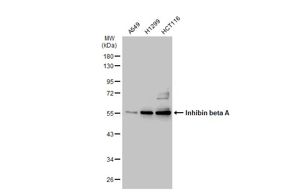
Inhibin beta A antibodyGTX108405
ApplicationsImmunoFluorescence, Western Blot, ImmunoCytoChemistry, ImmunoHistoChemistry, ImmunoHistoChemistry Paraffin
ReactivityHuman
TargetINHBA
- SizePrice