


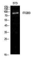
Figure 1. Western blot analysis of ITGB3 using anti-ITGB3 antibody (PA1627). Electrophoresis was performed on a 5-20% SDS-PAGE gel at 70V (Stacking gel) / 90V (Resolving gel) for 2-3 hours. The sample well of each lane was loaded with 30 ug of sample under reducing conditions. Lane 1: human U87 whole cell lysates, Lane 2: human HEL whole cell lysates, Lane 3: human Hela whole cell lysates, Lane 4: rat spleen tissue lysates, Lane 5: mouse spleen tissue lysates. After electrophoresis, proteins were transferred to a nitrocellulose membrane at 150 mA for 50-90 minutes. Blocked the membrane with 5% non-fat milk/TBS for 1.5 hour at RT. The membrane was incubated with rabbit anti-ITGB3 antigen affinity purified polyclonal antibody (Catalog # PA1627) at 0.5 microg/mL overnight at 4°C, then washed with TBS-0.1%Tween 3 times with 5 minutes each and probed with a goat anti-rabbit IgG-HRP secondary antibody at a dilution of 1:5000 for 1.5 hour at RT. The signal is developed using an Enhanced Chemiluminescent detection (ECL) kit (Catalog # EK1002) with Tanon 5200 system. A specific band was detected for ITGB3 at approximately 100 kDa. The expected band size for ITGB3 is at 87 kDa.
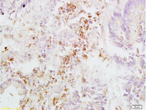
. ITGB3 was detected in a paraffin-embedded section of human laryngeal squamous cell carcinoma tissue. Heat mediated antigen retrieval was performed in EDTA buffer (pH 8.0, epitope retrieval solution). The tissue section was blocked with 10% goat serum. The tissue section was then incubated with 2 microg/ml rabbit anti-ITGB3 Antibody (PA1627) overnight at 4°C. Peroxidase Conjugated Goat Anti-rabbit IgG was used as secondary antibody and incubated for 30 minutes at 37°C. The tissue section was developed using HRP Conjugated Rabbit IgG Super Vision Assay Kit (Catalog # SV0002) with DAB as the chromogen.")
. ITGB3 was detected in a paraffin-embedded section of human liver cancer tissue. Heat mediated antigen retrieval was performed in EDTA buffer (pH 8.0, epitope retrieval solution). The tissue section was blocked with 10% goat serum. The tissue section was then incubated with 2 microg/ml rabbit anti-ITGB3 Antibody (PA1627) overnight at 4°C. Peroxidase Conjugated Goat Anti-rabbit IgG was used as secondary antibody and incubated for 30 minutes at 37°C. The tissue section was developed using HRP Conjugated Rabbit IgG Super Vision Assay Kit (Catalog # SV0002) with DAB as the chromogen.")
. ITGB3 was detected in a paraffin-embedded section of human thyroid cancer tissue. Heat mediated antigen retrieval was performed in EDTA buffer (pH 8.0, epitope retrieval solution). The tissue section was blocked with 10% goat serum. The tissue section was then incubated with 2 microg/ml rabbit anti-ITGB3 Antibody (PA1627) overnight at 4°C. Peroxidase Conjugated Goat Anti-rabbit IgG was used as secondary antibody and incubated for 30 minutes at 37°C. The tissue section was developed using HRP Conjugated Rabbit IgG Super Vision Assay Kit (Catalog # SV0002) with DAB as the chromogen.")
. ITGB3 was detected in a paraffin-embedded section of rat lung tissue. Heat mediated antigen retrieval was performed in EDTA buffer (pH 8.0, epitope retrieval solution). The tissue section was blocked with 10% goat serum. The tissue section was then incubated with 2 microg/ml rabbit anti-ITGB3 Antibody (PA1627) overnight at 4°C. Peroxidase Conjugated Goat Anti-rabbit IgG was used as secondary antibody and incubated for 30 minutes at 37°C. The tissue section was developed using HRP Conjugated Rabbit IgG Super Vision Assay Kit (Catalog # SV0002) with DAB as the chromogen.")
Figure 1. Western blot analysis of ITGB3 using anti-ITGB3 antibody (PA1627). Electrophoresis was performed on a 5-20% SDS-PAGE gel at 70V (Stacking gel) / 90V (Resolving gel) for 2-3 hours. The sample well of each lane was loaded with 30 ug of sample under reducing conditions. Lane 1: human U87 whole cell lysates, Lane 2: human HEL whole cell lysates, Lane 3: human Hela whole cell lysates, Lane 4: rat spleen tissue lysates, Lane 5: mouse spleen tissue lysates. After electrophoresis, proteins were transferred to a nitrocellulose membrane at 150 mA for 50-90 minutes. Blocked the membrane with 5% non-fat milk/TBS for 1.5 hour at RT. The membrane was incubated with rabbit anti-ITGB3 antigen affinity purified polyclonal antibody (Catalog # PA1627) at 0.5 microg/mL overnight at 4°C, then washed with TBS-0.1%Tween 3 times with 5 minutes each and probed with a goat anti-rabbit IgG-HRP secondary antibody at a dilution of 1:5000 for 1.5 hour at RT. The signal is developed using an Enhanced Chemiluminescent detection (ECL) kit (Catalog # EK1002) with Tanon 5200 system. A specific band was detected for ITGB3 at approximately 100 kDa. The expected band size for ITGB3 is at 87 kDa.
Anti-Integrin Beta 3 Antibody
PA1627
ApplicationsWestern Blot, ImmunoHistoChemistry
Product group Antibodies
ReactivityHuman, Mouse, Rat
TargetITGB3
Overview
- SupplierBoster Bio
- Product NameAnti-Integrin Beta 3 Antibody
- Delivery Days Customer9
- Application Supplier NoteTested Species: In-house tested species with positive results. Predicted Species: Species predicted to be fit for the product based on sequence similarities. By Heat: Boiling the paraffin sections in 10mM citrate buffer, pH6.0, for 20mins is required for the staining of formalin/paraffin sections. Other applications have not been tested. Optimal dilutions should be determined by end users.
- ApplicationsWestern Blot, ImmunoHistoChemistry
- Applications SupplierIHP, WB, IHC
- CertificationResearch Use Only
- ClonalityPolyclonal
- Concentration500 ug/ml
- Gene ID3690
- Target nameITGB3
- Target descriptionintegrin subunit beta 3
- Target synonymsBDPLT16, BDPLT2, BDPLT24, CD61, FMAIT1, GP3A, GPIIIa, GT, GT2, integrin beta-3, antigen CD61, integrin beta 3, integrin beta chain, beta 3, integrin, beta 3 (platelet glycoprotein IIIa, antigen CD61), platelet membrane glycoprotein IIIa
- HostRabbit
- IsotypeIgG
- Protein IDP05106
- Protein NameIntegrin beta-3
- Scientific DescriptionBoster Bio Anti-Integrin beta 3/ITGB3 Antibody catalog # PA1627. Tested in IHC, WB applications. This antibody reacts with Human, Mouse, Rat. The brand Picoband indicates this is a premium antibody that guarantees superior quality, high affinity, and strong signals with minimal background in Western blot applications. Only our best-performing antibodies are designated as Picoband, ensuring unmatched performance.
- ReactivityHuman, Mouse, Rat
- Reactivity SupplierHuman, Mouse, Rat, Hamster
- Storage Instruction-20°C,2°C to 8°C
- UNSPSC12352203
References
- Qin X, Yan M, Zhang J, et al. TGFβ3-mediated induction of Periostin facilitates head and neck cancer growth and is associated with metastasis. Sci Rep. 2016,6:20587. doi: 10.1038/srep20587Read this paper
- Wang Y, Liu J, Lin B, et al. Study on the expression and clinical significances of lewis y antigen and integrin αv, β3 in epithelial ovarian tumors. Int J Mol Sci. 2011,12(6):3409-21. doi: 10.3390/ijms12063409Read this paper
- Wang C, Yan L, Wang Y, et al. Overexpression of Lewis(y) antigen protects ovarian cancer RMG-1 cells from carboplatin-induced apoptosis by the upregulation of Topo-I and Topo-II β. Anat Rec (Hoboken). 2011,294(6):961-9. doi: 10.1002/ar.21398Read this paper
Datasheet
MSDS
Related products
Product group Antibodies
ReactivityHuman
TargetITGB3
- SizePrice
Product group Antibodies
ApplicationsFlow Cytometry
TargetITGB3
- SizePrice

Product group Antibodies
References
ITGB3 Polyclonal AntibodyBS-0342R
ApplicationsFlow Cytometry, ImmunoFluorescence, Western Blot, ELISA, ImmunoCytoChemistry, ImmunoHistoChemistry, ImmunoHistoChemistry Frozen, ImmunoHistoChemistry Paraffin
ReactivityBovine, Human, Mouse, Porcine, Rabbit
TargetITGB3
- SizePrice
Product group Antibodies
Anti-ITGB3 AntibodyA101008
ApplicationsWestern Blot, ELISA
ReactivityHuman
- SizePrice
Product group Antibodies
Anti-ITGB3 Antibody144-61563
ApplicationsWestern Blot
ReactivityHuman, Mouse, Rat
TargetITGB3
- SizePrice

Product group Antibodies
Anti-ITGB3 AntibodyAMAB91470
ApplicationsWestern Blot, ImmunoHistoChemistry
ReactivityHuman
TargetITGB3
- SizePrice
Product group Antibodies

Anti-Integrin beta-3 [Y2/51]Ab01334-1.1
ApplicationsFlow Cytometry, ImmunoFluorescence, Western Blot, ImmunoHistoChemistry
ReactivityHuman
TargetITGB3
- SizePrice
Product group Antibodies
Integrin beta 3 antibody [ZM33]GTX01641
ApplicationsImmunoHistoChemistry, ImmunoHistoChemistry Paraffin
ReactivityHuman
TargetITGB3
- SizePrice