

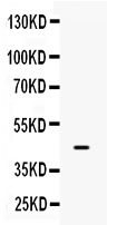
Figure 1. Western blot analysis of IRF9 using anti-IRF9 antibody (A04485). Electrophoresis was performed on a 5-20% SDS-PAGE gel at 70V (Stacking gel) / 90V (Resolving gel) for 2-3 hours. The sample well of each lane was loaded with 50ug of sample under reducing conditions. lane 1: JURKAT cell lysates. After Electrophoresis, proteins were transferred to a Nitrocellulose membrane at 150mA for 50-90 minutes. Blocked the membrane with 5% Non-fat Milk/ TBS for 1.5 hour at RT. The membrane was incubated with rabbit anti-IRF9 antigen affinity purified polyclonal antibody (Catalog # A04485) at 0.5 microg/mL overnight at 4°C, then washed with TBS-0.1%Tween 3 times with 5 minutes each and probed with a goat anti-rabbit IgG-HRP secondary antibody at a dilution of 1:10000 for 1.5 hour at RT. The signal is developed using an Enhanced Chemiluminescent detection (ECL) kit (Catalog # EK1002) with Tanon 5200 system. A specific band was detected for IRF9 at approximately 44KD. The expected band size for IRF9 is at 44KD.
Figure 1. Western blot analysis of IRF9 using anti-IRF9 antibody (A04485). Electrophoresis was performed on a 5-20% SDS-PAGE gel at 70V (Stacking gel) / 90V (Resolving gel) for 2-3 hours. The sample well of each lane was loaded with 50ug of sample under reducing conditions. lane 1: JURKAT cell lysates. After Electrophoresis, proteins were transferred to a Nitrocellulose membrane at 150mA for 50-90 minutes. Blocked the membrane with 5% Non-fat Milk/ TBS for 1.5 hour at RT. The membrane was incubated with rabbit anti-IRF9 antigen affinity purified polyclonal antibody (Catalog # A04485) at 0.5 microg/mL overnight at 4°C, then washed with TBS-0.1%Tween 3 times with 5 minutes each and probed with a goat anti-rabbit IgG-HRP secondary antibody at a dilution of 1:10000 for 1.5 hour at RT. The signal is developed using an Enhanced Chemiluminescent detection (ECL) kit (Catalog # EK1002) with Tanon 5200 system. A specific band was detected for IRF9 at approximately 44KD. The expected band size for IRF9 is at 44KD.
Anti-Interferon regulatory factor 9/IRF9 Antibody Picoband(r)
A04485-CARRIER-FREE
ApplicationsFlow Cytometry, ImmunoFluorescence, Western Blot, ImmunoCytoChemistry, ImmunoHistoChemistry
Product group Antibodies
ReactivityHuman
TargetIRF9
Overview
- SupplierBoster Bio
- Product NameAnti-Interferon regulatory factor 9/IRF9 Antibody Picoband(r)
- Delivery Days Customer9
- Application Supplier NoteTested Species: In-house tested species with positive results. Other applications have not been tested. Optimal dilutions should be determined by end users.
- ApplicationsFlow Cytometry, ImmunoFluorescence, Western Blot, ImmunoCytoChemistry, ImmunoHistoChemistry
- CertificationResearch Use Only
- ClonalityPolyclonal
- Concentration500 ug/ml
- Gene ID10379
- Target nameIRF9
- Target descriptioninterferon regulatory factor 9
- Target synonymsIRF-9, ISGF3, ISGF3G, p48, interferon regulatory factor 9, IFN-alpha-responsive transcription factor subunit, ISGF-3 gamma, ISGF3 p48 subunit, interferon-stimulated gene factor 3 gamma, interferon-stimulated transcription factor 3, gamma (48kD), interferon-stimulated transcription factor 3, gamma 48kDa, transcriptional regulator ISGF3 subunit gamma
- HostRabbit
- IsotypeIgG
- Scientific DescriptionBoster Bio Anti-Interferon regulatory factor 9/IRF9 Antibody Picoband® catalog # A04485. Tested in Flow Cytometry, IF, IHC, ICC, WB applications. This antibody reacts with Human. The brand Picoband indicates this is a premium antibody that guarantees superior quality, high affinity, and strong signals with minimal background in Western blot applications. Only our best-performing antibodies are designated as Picoband, ensuring unmatched performance.
- ReactivityHuman
- Storage Instruction-20°C,2°C to 8°C
- UNSPSC12352203
Related products
Product group Antibodies
Anti-IRF9 AntibodyA38674
ApplicationsWestern Blot, ImmunoHistoChemistry
ReactivityHuman, Mouse
- SizePrice
Product group Antibodies
IRF9/ISGF3 Recombinant Antibody, AbBy Fluor-647 ConjugatedBSM-62012R-BF647
ApplicationsWestern Blot
ReactivityHuman
TargetIRF9
- SizePrice
Product group Antibodies
IRF9 AntibodyCSB-PA011824LA01HU
ApplicationsImmunoFluorescence, Western Blot, ELISA
ReactivityHuman, Mouse
TargetIRF9
- SizePrice
Product group Antibodies
IRF9 Polyclonal AntibodyCAC15050
ApplicationsImmunoFluorescence, Western Blot, ELISA
ReactivityMouse
TargetIRF9
- SizePrice

Product group Antibodies
Anti-IRF9 AntibodyHPA001862
ApplicationsImmunoCytoChemistry, ImmunoHistoChemistry
ReactivityHuman
TargetIRF9
- SizePrice
Product group Antibodies
IRF9 antibodyGTX115401
ApplicationsWestern Blot, ImmunoHistoChemistry, ImmunoHistoChemistry Paraffin
ReactivityHuman
TargetIRF9
- SizePrice
Product group Antibodies
Anti-IRF9Y058880
ApplicationsWestern Blot, ELISA, ImmunoHistoChemistry
ReactivityHuman
- SizePrice
Product group Antibodies

ISGF3 / IRF9 AntibodyLS-C404084
ApplicationsWestern Blot, ELISA, ImmunoHistoChemistry
ReactivityHuman, Mouse
TargetIRF9
- SizePrice