


Immunohistochemical staining of human pancreas shows strong cytoplasmic positivity in exocrine glands.

Immunohistochemical staining of human pancreas shows strong cytoplasmic positivity in exocrine glands.
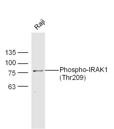
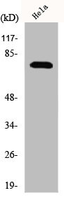
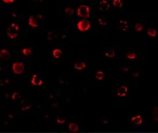
Anti-IRAK1 Antibody
HPA054476
ApplicationsImmunoCytoChemistry, ImmunoHistoChemistry
Product group Antibodies
ReactivityHuman
TargetIRAK1
Overview
- SupplierAtlas Antibodies
- Product NameAnti-IRAK1 Antibody
- Delivery Days Customer4
- ApplicationsImmunoCytoChemistry, ImmunoHistoChemistry
- CertificationResearch Use Only
- ClonalityPolyclonal
- ConjugateUnconjugated
- Gene ID3654
- Target nameIRAK1
- Target descriptioninterleukin 1 receptor associated kinase 1
- Target synonymsIRAK, pelle, interleukin-1 receptor-associated kinase 1, Pelle homolog
- HostRabbit
- IsotypeIgG
- Protein IDP51617
- Protein NameInterleukin-1 receptor-associated kinase 1
- Scientific DescriptionRecombinant Protein Epitope Signature Tag (PrEST) antigen sequence
- ReactivityHuman
- Storage Instruction-20°C,2°C to 8°C
- UNSPSC41116161
Datasheet
MSDS
Related products
Product group Antibodies

IRAK1 (Phospho-Ser376) AntibodyABX012604
ApplicationsELISA, ImmunoHistoChemistry
- SizePrice

Product group Antibodies
Anti-IRAK1 AntibodyA100667
ApplicationsELISA, ImmunoHistoChemistry
ReactivityHuman
- SizePrice

Product group Antibodies
Anti-IRAK-1/IRAK1 Antibody Picoband(r)A01021-1-CARRIER-FREE
ApplicationsFlow Cytometry, ImmunoFluorescence, Western Blot, ImmunoCytoChemistry, ImmunoHistoChemistry
ReactivityHuman, Mouse, Rat
TargetIRAK1
- SizePrice
Product group Antibodies
Anti-IRAK1 Antibody144-12624
ApplicationsImmunoFluorescence, Western Blot
ReactivityHuman, Mouse, Rat
TargetIRAK1
- SizePrice
Product group Antibodies

IRAK1 / IRAK AntibodyLS-C747718
ApplicationsImmunoFluorescence, Western Blot
ReactivityHuman, Mouse, Rat
TargetIRAK1
- SizePrice
Product group Antibodies
ApplicationsFlow Cytometry, ImmunoFluorescence, Western Blot, ImmunoCytoChemistry, ImmunoHistoChemistry, ImmunoHistoChemistry Frozen, ImmunoHistoChemistry Paraffin
ReactivityHuman, Mouse, Rat
TargetIRAK1
- SizePrice
Product group Antibodies
IRAK1 AntibodyCSB-PA003061
ApplicationsImmunoFluorescence, Western Blot, ELISA
ReactivityHuman, Mouse, Rat
TargetIRAK1
- SizePrice
Product group Antibodies
ApplicationsImmunoPrecipitation, Western Blot, ImmunoCytoChemistry, ImmunoHistoChemistry
ReactivityMouse, Rat
TargetIRAK1
- SizePrice
Product group Antibodies
References
IRAK1 antibodyGTX31253
ApplicationsImmunoFluorescence, ImmunoPrecipitation, Western Blot, ELISA, ImmunoCytoChemistry
ReactivityHuman, Mouse, Rat
TargetIRAK1
- SizePrice