


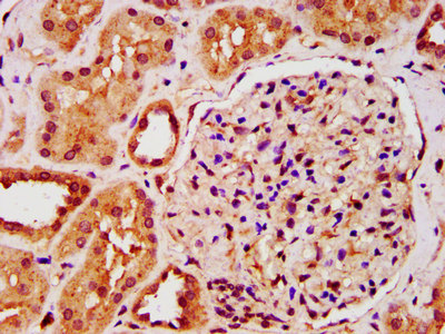
Immunohistochemical staining of human placenta shows strong cytoplasmic positivity in trophoblastic cells.
 shows similar pattern to independent antibody HPA054532 (B).")
Immunohistochemical staining of human placenta shows strong cytoplasmic positivity in trophoblastic cells.
Anti-IST1 Antibody
HPA041802
ApplicationsWestern Blot, ImmunoHistoChemistry
Product group Antibodies
ReactivityHuman
TargetIST1
Overview
- SupplierAtlas Antibodies
- Product NameAnti-IST1 Antibody
- Delivery Days Customer4
- ApplicationsWestern Blot, ImmunoHistoChemistry
- CertificationResearch Use Only
- ClonalityPolyclonal
- ConjugateUnconjugated
- Gene ID9798
- Target nameIST1
- Target descriptionIST1 factor associated with ESCRT-III
- Target synonymsCHMP8, OLC1, IST1 homolog, IST1, ESCRT-III associated factor, IST1, endosomal sorting complex required for transport-III component, charged multivesicular body protein 8, increased sodium tolerance 1 homolog, overexpressed in lung cancer 1, putative MAPK-activating protein PM28
- HostRabbit
- IsotypeIgG
- Protein IDP53990
- Protein NameIST1 homolog
- Scientific DescriptionRecombinant Protein Epitope Signature Tag (PrEST) antigen sequence
- ReactivityHuman
- Storage Instruction-20°C,2°C to 8°C
- UNSPSC41116161
Datasheet
MSDS
Related products
Product group Antibodies
IST1 AntibodyCSB-PA012171LA01HU
ApplicationsImmunoFluorescence, ELISA, ImmunoHistoChemistry
ReactivityHuman
TargetIST1
- SizePrice

Product group Antibodies
Anti-hIST1/IST1 Antibody Picoband(r)A05507-2-CARRIER-FREE
ApplicationsFlow Cytometry, ImmunoFluorescence, Western Blot, ELISA, ImmunoCytoChemistry, ImmunoHistoChemistry
ReactivityHuman, Mouse, Rat
TargetIST1
- SizePrice
Product group Antibodies

IST1 AntibodyLS-C830187
ApplicationsWestern Blot, ELISA, ImmunoHistoChemistry
ReactivityHuman, Mouse, Rat
TargetIST1
- SizePrice

Product group Antibodies
Anti-IST1 AntibodyHPA054532
ApplicationsWestern Blot, ImmunoCytoChemistry, ImmunoHistoChemistry
ReactivityHuman
TargetIST1
- SizePrice
Product group Antibodies
Anti-IST1 AntibodyHPA054532
ApplicationsWestern Blot, ImmunoCytoChemistry, ImmunoHistoChemistry
ReactivityHuman
TargetIST1
- SizePrice
Product group Antibodies
IST1 antibodyGTX101972
ApplicationsWestern Blot
ReactivityHuman, Mouse
TargetIST1
- SizePrice
Product group Antibodies
Anti-IST1 Antibody144-61474
ApplicationsWestern Blot
ReactivityHuman, Mouse, Rat
TargetIST1
- SizePrice