


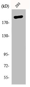
Figure 1. Western blot analysis of KAT3B/P300/EP300 using anti-KAT3B/P300/EP300 antibody (A00117). Electrophoresis was performed on a 5-20% SDS-PAGE gel at 70V (Stacking gel) / 90V (Resolving gel) for 2-3 hours. The sample well of each lane was loaded with 30 ug of sample under reducing conditions. Lane 1: human 293T whole cell lysates, Lane 2: mouse testis tissue lysates. After electrophoresis, proteins were transferred to a nitrocellulose membrane at 150 mA for 50-90 minutes. Blocked the membrane with 5% non-fat milk/TBS for 1.5 hour at RT. The membrane was incubated with rabbit anti-KAT3B/P300/EP300 antigen affinity purified polyclonal antibody (Catalog # A00117) at 0.5 microg/mL overnight at 4°C, then washed with TBS-0.1%Tween 3 times with 5 minutes each and probed with a goat anti-rabbit IgG-HRP secondary antibody at a dilution of 1:5000 for 1.5 hour at RT. The signal is developed using an Enhanced Chemiluminescent detection (ECL) kit (Catalog # EK1002) with Tanon 5200 system. A specific band was detected for KAT3B/P300/EP300 at approximately 264-300 kDa. The expected band size for KAT3B/P300/EP300 is at 264 kDa.
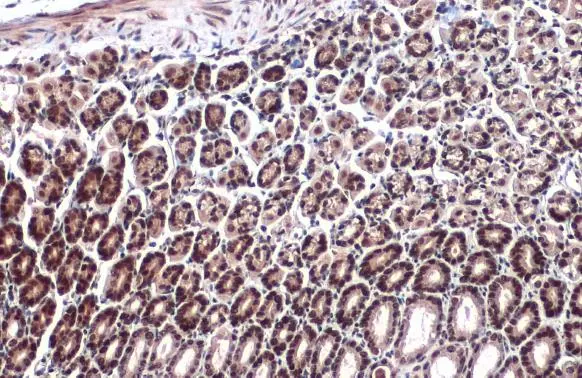
. KAT3B/P300/EP300 was detected in a paraffin-embedded section of differentiated adenocarcinoma of the human rectum tissue. Heat mediated antigen retrieval was performed in EDTA buffer (pH 8.0, epitope retrieval solution). The tissue section was blocked with 10% goat serum. The tissue section was then incubated with 2 microg/ml rabbit anti-KAT3B/P300/EP300 Antibody (A00117) overnight at 4°C. Biotinylated goat anti-rabbit IgG was used as secondary antibody and incubated for 30 minutes at 37°C. The tissue section was developed using Strepavidin-Biotin-Complex (SABC) (Catalog # SA1022) with DAB as the chromogen.")
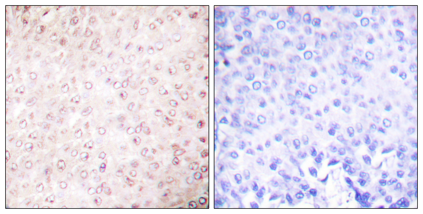
. KAT3B/P300/EP300 was detected in a paraffin-embedded section of human classic Hodgkins lymphoma tissue. Heat mediated antigen retrieval was performed in EDTA buffer (pH 8.0, epitope retrieval solution). The tissue section was blocked with 10% goat serum. The tissue section was then incubated with 2 microg/ml rabbit anti-KAT3B/P300/EP300 Antibody (A00117) overnight at 4°C. Biotinylated goat anti-rabbit IgG was used as secondary antibody and incubated for 30 minutes at 37°C. The tissue section was developed using Strepavidin-Biotin-Complex (SABC) (Catalog # SA1022) with DAB as the chromogen.")
. KAT3B/P300/EP300 was detected in a paraffin-embedded section of human gastric signet ring cell carcinoma tissue. Heat mediated antigen retrieval was performed in EDTA buffer (pH 8.0, epitope retrieval solution). The tissue section was blocked with 10% goat serum. The tissue section was then incubated with 2 microg/ml rabbit anti-KAT3B/P300/EP300 Antibody (A00117) overnight at 4°C. Biotinylated goat anti-rabbit IgG was used as secondary antibody and incubated for 30 minutes at 37°C. The tissue section was developed using Strepavidin-Biotin-Complex (SABC) (Catalog # SA1022) with DAB as the chromogen.")
. KAT3B/P300/EP300 was detected in a paraffin-embedded section of human placenta tissue. Heat mediated antigen retrieval was performed in EDTA buffer (pH 8.0, epitope retrieval solution). The tissue section was blocked with 10% goat serum. The tissue section was then incubated with 2 microg/ml rabbit anti-KAT3B/P300/EP300 Antibody (A00117) overnight at 4°C. Biotinylated goat anti-rabbit IgG was used as secondary antibody and incubated for 30 minutes at 37°C. The tissue section was developed using Strepavidin-Biotin-Complex (SABC) (Catalog # SA1022) with DAB as the chromogen.")
. KAT3B/P300/EP300 was detected in an immunocytochemical section of SiHa cells. Enzyme antigen retrieval was performed using IHC enzyme antigen retrieval reagent (AR0022) for 15 mins. The cells were blocked with 10% goat serum. And then incubated with 5 microg/mL rabbit anti-KAT3B/P300/EP300 Antibody (A00117) overnight at 4°C. DyLight®488 Conjugated Goat Anti-Rabbit IgG (BA1127) was used as secondary antibody at 1:100 dilution and incubated for 30 minutes at 37°C. The section was counterstained with DAPI. Visualize using a fluorescence microscope and filter sets appropriate for the label used.")
. Overlay histogram showing 293T cells stained with A00117 (Blue line). To facilitate intracellular staining, cells were fixed with 4% paraformaldehyde and permeabilized with permeabilization buffer. The cells were blocked with 10% normal goat serum. And then incubated with rabbit anti-KAT3B/P300/EP300 Antibody (A00117, 1 microg/1x106 cells) for 30 min at 20°C. DyLight®488 conjugated goat anti-rabbit IgG (BA1127, 5-10 microg/1x106 cells) was used as secondary antibody for 30 minutes at 20°C. Isotype control antibody (Green line) was rabbit IgG (1 microg/1x106) used under the same conditions. Unlabelled sample without incubation with primary antibody and secondary antibody (Red line) was used as a blank control.")
Figure 1. Western blot analysis of KAT3B/P300/EP300 using anti-KAT3B/P300/EP300 antibody (A00117). Electrophoresis was performed on a 5-20% SDS-PAGE gel at 70V (Stacking gel) / 90V (Resolving gel) for 2-3 hours. The sample well of each lane was loaded with 30 ug of sample under reducing conditions. Lane 1: human 293T whole cell lysates, Lane 2: mouse testis tissue lysates. After electrophoresis, proteins were transferred to a nitrocellulose membrane at 150 mA for 50-90 minutes. Blocked the membrane with 5% non-fat milk/TBS for 1.5 hour at RT. The membrane was incubated with rabbit anti-KAT3B/P300/EP300 antigen affinity purified polyclonal antibody (Catalog # A00117) at 0.5 microg/mL overnight at 4°C, then washed with TBS-0.1%Tween 3 times with 5 minutes each and probed with a goat anti-rabbit IgG-HRP secondary antibody at a dilution of 1:5000 for 1.5 hour at RT. The signal is developed using an Enhanced Chemiluminescent detection (ECL) kit (Catalog # EK1002) with Tanon 5200 system. A specific band was detected for KAT3B/P300/EP300 at approximately 264-300 kDa. The expected band size for KAT3B/P300/EP300 is at 264 kDa.
Anti-KAT3B/p300/EP300 Antibody Picoband(r)
A00117-CARRIER-FREE
ApplicationsFlow Cytometry, ImmunoFluorescence, Western Blot, ImmunoCytoChemistry, ImmunoHistoChemistry
Product group Antibodies
ReactivityHuman, Mouse
TargetEP300
Overview
- SupplierBoster Bio
- Product NameAnti-KAT3B/p300/EP300 Antibody Picoband(r)
- Delivery Days Customer9
- Application Supplier NoteTested Species: In-house tested species with positive results. Other applications have not been tested. Optimal dilutions should be determined by end users.
- ApplicationsFlow Cytometry, ImmunoFluorescence, Western Blot, ImmunoCytoChemistry, ImmunoHistoChemistry
- CertificationResearch Use Only
- ClonalityPolyclonal
- Concentration500 ug/ml
- Gene ID2033
- Target nameEP300
- Target descriptionEP300 lysine acetyltransferase
- Target synonymsKAT3B, MKHK2, RSTS2, p300, histone acetyltransferase p300, E1A binding protein p300, E1A-associated protein p300, E1A-binding protein, 300kD, histone butyryltransferase p300, histone crotonyltransferase p300, p300 HAT, protein 2-hydroxyisobutyryltransferase p300, protein lactyltransferas p300, protein propionyltransferase p300
- HostRabbit
- IsotypeIgG
- Protein IDQ09472
- Protein NameHistone acetyltransferase p300
- Scientific DescriptionBoster Bio Anti-KAT3B/p300/EP300 Antibody Picoband® catalog # A00117. Tested in Flow Cytometry, IF, IHC, ICC, WB applications. This antibody reacts with Human, Mouse. The brand Picoband indicates this is a premium antibody that guarantees superior quality, high affinity, and strong signals with minimal background in Western blot applications. Only our best-performing antibodies are designated as Picoband, ensuring unmatched performance.
- ReactivityHuman, Mouse
- Storage Instruction-20°C,2°C to 8°C
- UNSPSC12352203
Related products
Product group Antibodies
Anti-p300 AntibodyA96102
ApplicationsWestern Blot, ELISA, ImmunoHistoChemistry
ReactivityHuman, Mouse, Rat
- SizePrice
Product group Antibodies

Anti-EP300 [RAB-C318]Ab01774-1.1
ApplicationsImmunoFluorescence, ImmunoPrecipitation
ReactivityHuman
TargetEP300
- SizePrice
Product group Antibodies
Anti-EP300 Antibody144-62588
ApplicationsImmunoFluorescence, Western Blot, ImmunoHistoChemistry
ReactivityHuman, Mouse, Rat
TargetEP300
- SizePrice

Product group Antibodies
References
ApplicationsImmunoFluorescence, Western Blot, ELISA, ImmunoCytoChemistry, ImmunoHistoChemistry, ImmunoHistoChemistry Frozen, ImmunoHistoChemistry Paraffin
ReactivityBovine, Canine, Chicken, Equine, Guinea Pig, Human, Mouse, Porcine, Rat
TargetEP300
- SizePrice
Product group Antibodies
EP300 AntibodyCSB-PA003640
ApplicationsWestern Blot, ELISA, ImmunoHistoChemistry
ReactivityHuman, Mouse, Rat
TargetEP300
- SizePrice
Product group Antibodies
ApplicationsImmunoPrecipitation, Western Blot, ImmunoCytoChemistry, ImmunoHistoChemistry
ReactivityPorcine
TargetEP300
- SizePrice
Product group Antibodies
EP300 antibodyGTX134238
ApplicationsWestern Blot, ImmunoHistoChemistry, ImmunoHistoChemistry Paraffin
ReactivityHuman, Mouse
TargetEP300
- SizePrice
Product group Antibodies

EP300 / p300 Antibody (Internal)LS-C368790
ApplicationsWestern Blot
ReactivityHuman, Mouse
TargetEP300
- SizePrice

Product group Antibodies
Anti-EP300 AntibodyHPA003128
ApplicationsChIP Chromatin ImmunoPrecipitation, ImmunoCytoChemistry, ImmunoHistoChemistry
ReactivityHuman
TargetEP300
- SizePrice