


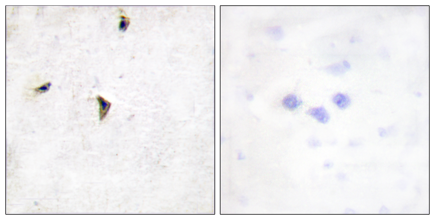
Immunohistochemical staining of human cerebral cortex shows strong membranous positivity in neurons.
Immunohistochemical staining of human cerebral cortex shows strong membranous positivity in neurons.
Anti-KCNB1 Antibody
HPA042434
ApplicationsImmunoHistoChemistry
Product group Antibodies
ReactivityHuman
TargetKCNB1
Overview
- SupplierAtlas Antibodies
- Product NameAnti-KCNB1 Antibody
- Delivery Days Customer4
- ApplicationsImmunoHistoChemistry
- CertificationResearch Use Only
- ClonalityPolyclonal
- ConjugateUnconjugated
- Gene ID3745
- Target nameKCNB1
- Target descriptionpotassium voltage-gated channel subfamily B member 1
- Target synonymsDEE26, DRK1, Kv2.1, potassium voltage-gated channel subfamily B member 1, delayed rectifier potassium channel 1, potassium voltage-gated channel, Shab-related subfamily, member 1, voltage-gated potassium channel subunit Kv2.1
- HostRabbit
- IsotypeIgG
- Protein IDQ14721
- Protein NamePotassium voltage-gated channel subfamily B member 1
- Scientific DescriptionRecombinant Protein Epitope Signature Tag (PrEST) antigen sequence
- ReactivityHuman
- Storage Instruction-20°C,2°C to 8°C
- UNSPSC41116161
Datasheet
MSDS
Related products
Product group Antibodies
Anti-KCNB1 AntibodyA95756
ApplicationsImmunoFluorescence, ELISA, ImmunoHistoChemistry
ReactivityHuman, Mouse, Rat
- SizePrice
Product group Antibodies

Anti-Kv2.1 K+ Channel Ectodomain [K39/25]Ab02110-10.0
ApplicationsImmunoFluorescence, ImmunoPrecipitation, Western Blot
ReactivityHuman, Mouse, Rat
TargetKCNB1
- SizePrice
Product group Antibodies

Kv2.1 (Phospho-Ser805) AntibodyABX012695
ApplicationsWestern Blot, ELISA
- SizePrice
Product group Antibodies
Anti-Kv2.1 (Phospho-Ser805) Antibody102-26398
ApplicationsWestern Blot
TargetKCNB1
- SizePrice
Product group Antibodies

KCNB1 / Kv2.1 AntibodyLS-C813447
ApplicationsWestern Blot, ELISA
ReactivityHuman, Mouse, Rat
TargetKCNB1
- SizePrice
Product group Antibodies
ApplicationsImmunoFluorescence, ELISA, ImmunoCytoChemistry, ImmunoHistoChemistry, ImmunoHistoChemistry Frozen, ImmunoHistoChemistry Paraffin
ReactivityBovine, Canine, Equine, Human, Mouse, Porcine, Rat, Sheep
TargetKCNB1
- SizePrice
Product group Antibodies
KCNB1 AntibodyCSB-PA010500
ApplicationsWestern Blot, ELISA
ReactivityHuman, Mouse, Rat
TargetKCNB1
- SizePrice
Product group Antibodies

Goat anti-KCNB1 / DRK1EB08061
ApplicationsELISA
ReactivityBovine, Canine, Human, Mouse, Porcine
TargetKCNB1
- SizePrice