


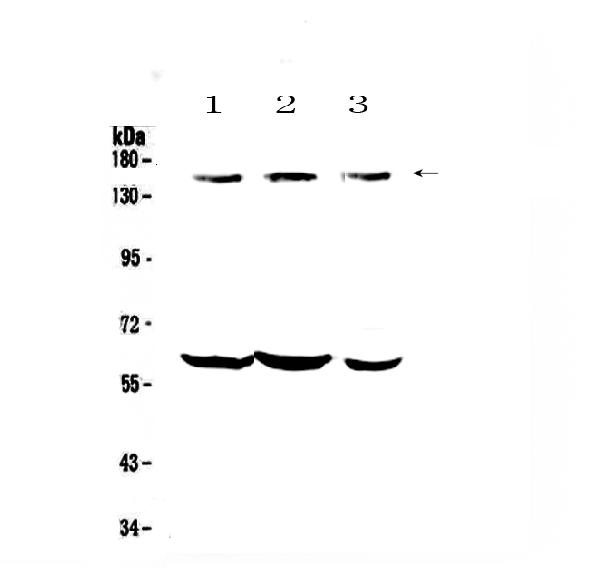
Figure 1. Western blot analysis of KCNH1 using anti-KCNH1 antibody (A01036-4). Electrophoresis was performed on a 5-20% SDS-PAGE gel at 70V (Stacking gel) / 90V (Resolving gel) for 2-3 hours. The sample well of each lane was loaded with 30 ug of sample under reducing conditions. Lane 1: human U-87MG whole cell lysates, Lane 2: human SH-SY5Y whole cell lysates, Lane 3: human Hela whole cell lysates, Lane 4: human MCF-7 whole cell lysates, Lane 5: rat brain tissue lysates, Lane 6: rat C6 whole cell lysates. After electrophoresis, proteins were transferred to a nitrocellulose membrane at 150 mA for 50-90 minutes. Blocked the membrane with 5% non-fat milk/TBS for 1.5 hour at RT. The membrane was incubated with rabbit anti-KCNH1 antigen affinity purified polyclonal antibody (Catalog # A01036-4) at 0.5 microg/mL overnight at 4°C, then washed with TBS-0.1%Tween 3 times with 5 minutes each and probed with a goat anti-rabbit IgG-HRP secondary antibody at a dilution of 1:5000 for 1.5 hour at RT. The signal is developed using an Enhanced Chemiluminescent detection (ECL) kit (Catalog # EK1002) with Tanon 5200 system. A specific band was detected for KCNH1 at approximately 111 kDa. The expected band size for KCNH1 is at 111 kDa.
Figure 1. Western blot analysis of KCNH1 using anti-KCNH1 antibody (A01036-4). Electrophoresis was performed on a 5-20% SDS-PAGE gel at 70V (Stacking gel) / 90V (Resolving gel) for 2-3 hours. The sample well of each lane was loaded with 30 ug of sample under reducing conditions. Lane 1: human U-87MG whole cell lysates, Lane 2: human SH-SY5Y whole cell lysates, Lane 3: human Hela whole cell lysates, Lane 4: human MCF-7 whole cell lysates, Lane 5: rat brain tissue lysates, Lane 6: rat C6 whole cell lysates. After electrophoresis, proteins were transferred to a nitrocellulose membrane at 150 mA for 50-90 minutes. Blocked the membrane with 5% non-fat milk/TBS for 1.5 hour at RT. The membrane was incubated with rabbit anti-KCNH1 antigen affinity purified polyclonal antibody (Catalog # A01036-4) at 0.5 microg/mL overnight at 4°C, then washed with TBS-0.1%Tween 3 times with 5 minutes each and probed with a goat anti-rabbit IgG-HRP secondary antibody at a dilution of 1:5000 for 1.5 hour at RT. The signal is developed using an Enhanced Chemiluminescent detection (ECL) kit (Catalog # EK1002) with Tanon 5200 system. A specific band was detected for KCNH1 at approximately 111 kDa. The expected band size for KCNH1 is at 111 kDa.
Anti-KCNH1 Antibody Picoband(r)
A01036-4-CARRIER-FREE
ApplicationsWestern Blot
Product group Antibodies
ReactivityHuman, Rat
TargetKCNH1
Overview
- SupplierBoster Bio
- Product NameAnti-KCNH1 Antibody Picoband(r)
- Delivery Days Customer9
- ApplicationsWestern Blot
- CertificationResearch Use Only
- ClonalityPolyclonal
- Concentration500 ug/ml
- Gene ID3756
- Target nameKCNH1
- Target descriptionpotassium voltage-gated channel subfamily H member 1
- Target synonymsEAG, EAG1, K(V)10.1, Kv10.1, TMBTS, ZLS1, h-eag, hEAG, hEAG1, voltage-gated delayed rectifier potassium channel KCNH1, EAG channel 1, ether-a-go-go 1, ether-a-go-go potassium channel 1, ether-a-go-go, Drosophila, homolog of, potassium channel, voltage gated eag related subfamily H, member 1, potassium voltage-gated channel, subfamily H (eag-related), member 1, voltage-gated potassium channel subunit Kv10.1
- HostRabbit
- IsotypeIgG
- Protein IDO95259
- Protein NameVoltage-gated delayed rectifier potassium channel KCNH1
- Scientific DescriptionBoster Bio Anti-KCNH1 Antibody Picoband® catalog # A01036-4. Tested in WB applications. This antibody reacts with Human, Rat. The brand Picoband indicates this is a premium antibody that guarantees superior quality, high affinity, and strong signals with minimal background in Western blot applications. Only our best-performing antibodies are designated as Picoband, ensuring unmatched performance.
- ReactivityHuman, Rat
- Storage Instruction-20°C,2°C to 8°C
- UNSPSC12352203
Related products
Product group Antibodies
KCNH1 Polyclonal AntibodyBS-2424R
ApplicationsFlow Cytometry, ImmunoFluorescence, Western Blot, ELISA, ImmunoCytoChemistry, ImmunoHistoChemistry, ImmunoHistoChemistry Frozen, ImmunoHistoChemistry Paraffin
ReactivityBovine, Canine, Equine, Human, Mouse, Porcine, Rabbit, Rat
TargetKCNH1
- SizePrice
Product group Antibodies
Anti-KCNH1 Antibody144-06636
ApplicationsWestern Blot
ReactivityHuman, Mouse, Rat
TargetKCNH1
- SizePrice
Product group Antibodies
Anti-KCNH1 AntibodyA97442
ApplicationsWestern Blot, ELISA
ReactivityHuman, Mouse, Rat
- SizePrice
Product group Antibodies
KCNH1 antibodyGTX64743
ApplicationsWestern Blot
ReactivityHuman
TargetKCNH1
- SizePrice
Product group Antibodies

Kv10.1 / KCNH1 AntibodyLS-C335693
ApplicationsWestern Blot, ImmunoHistoChemistry
ReactivityHuman, Mouse, Rat
TargetKCNH1
- SizePrice

Product group Antibodies
Anti-KCNH1 AntibodyHPA019445
ApplicationsImmunoHistoChemistry
ReactivityHuman
TargetKCNH1
- SizePrice
Product group Antibodies
Anti-KCNH1 Antibody Picoband(r)A01036-2-CARRIER-FREE
ApplicationsWestern Blot
ReactivityHuman, Mouse, Rat
TargetKCNH1
- SizePrice
Product group Antibodies
KCNH1 AntibodyCSB-PA003093
ApplicationsWestern Blot, ELISA
ReactivityHuman, Mouse, Rat
TargetKCNH1
- SizePrice