


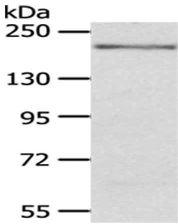
Figure 1. Western blot analysis of KDM6B using anti-KDM6B antibody (A01309-1). Electrophoresis was performed on a 5-20% SDS-PAGE gel at 70V (Stacking gel) / 90V (Resolving gel) for 2-3 hours. The sample well of each lane was loaded with 50ug of sample under reducing conditions. Lane 1: human K562 whole cell lysates, Lane 2: human A375 whole cell lysates, Lane 3: human HEK293 whole cell lysates, Lane 4: human A431 whole cell lysates. After Electrophoresis, proteins were transferred to a Nitrocellulose membrane at 150mA for 50-90 minutes. Blocked the membrane with 5% Non-fat Milk/ TBS for 1.5 hour at RT. The membrane was incubated with rabbit anti-KDM6B antigen affinity purified polyclonal antibody (Catalog # A01309-1) at 0.5 microg/mL overnight at 4°C, then washed with TBS-0.1%Tween 3 times with 5 minutes each and probed with a goat anti-rabbit IgG-HRP secondary antibody at a dilution of 1:5000 for 1.5 hour at RT. The signal is developed using an Enhanced Chemiluminescent detection (ECL) kit (Catalog # EK1002) with Tanon 5200 system. A specific band was detected for KDM6B at approximately 177KD. The expected band size for KDM6B is at 177KD.
. Overlay histogram showing K562 cells stained with A01309-1 (Blue line). To facilitate intracellular staining, cells were fixed with 4% paraformaldehyde and permeabilized with permeabilization buffer. The cells were blocked with 10% normal goat serum. And then incubated with rabbit anti-KDM6B Antibody (A01309-1, 1microg/1x106 cells) for 30 min at 20°C. DyLight®488 conjugated goat anti-rabbit IgG (BA1127, 5-10microg/1x106 cells) was used as secondary antibody for 30 minutes at 20°C. Isotype control antibody (Green line) was rabbit IgG (1microg/1x106) used under the same conditions. Unlabelled sample without incubation with primary antibody and secondary antibody (Red line) was used as a blank control.")
. Overlay histogram showing HEPA1-6 cells stained with A01309-1 (Blue line). To facilitate intracellular staining, cells were fixed with 4% paraformaldehyde and permeabilized with permeabilization buffer. The cells were blocked with 10% normal goat serum. And then incubated with rabbit anti-KDM6B Antibody (A01309-1, 1microg/1x106 cells) for 30 min at 20°C. DyLight®488 conjugated goat anti-rabbit IgG (BA1127, 5-10microg/1x106 cells) was used as secondary antibody for 30 minutes at 20°C. Isotype control antibody (Green line) was rabbit IgG (1microg/1x106) used under the same conditions. Unlabelled sample without incubation with primary antibody and secondary antibody (Red line) was used as a blank control.")
Figure 1. Western blot analysis of KDM6B using anti-KDM6B antibody (A01309-1). Electrophoresis was performed on a 5-20% SDS-PAGE gel at 70V (Stacking gel) / 90V (Resolving gel) for 2-3 hours. The sample well of each lane was loaded with 50ug of sample under reducing conditions. Lane 1: human K562 whole cell lysates, Lane 2: human A375 whole cell lysates, Lane 3: human HEK293 whole cell lysates, Lane 4: human A431 whole cell lysates. After Electrophoresis, proteins were transferred to a Nitrocellulose membrane at 150mA for 50-90 minutes. Blocked the membrane with 5% Non-fat Milk/ TBS for 1.5 hour at RT. The membrane was incubated with rabbit anti-KDM6B antigen affinity purified polyclonal antibody (Catalog # A01309-1) at 0.5 microg/mL overnight at 4°C, then washed with TBS-0.1%Tween 3 times with 5 minutes each and probed with a goat anti-rabbit IgG-HRP secondary antibody at a dilution of 1:5000 for 1.5 hour at RT. The signal is developed using an Enhanced Chemiluminescent detection (ECL) kit (Catalog # EK1002) with Tanon 5200 system. A specific band was detected for KDM6B at approximately 177KD. The expected band size for KDM6B is at 177KD.
Anti-KDM6B/JMJD3 Picoband(r) Antibody
A01309-1
ApplicationsFlow Cytometry, Western Blot, ELISA
Product group Antibodies
ReactivityHuman, Mouse
TargetKDM6B
Overview
- SupplierBoster Bio
- Product NameAnti-KDM6B/JMJD3 Picoband(r) Antibody
- Delivery Days Customer9
- ApplicationsFlow Cytometry, Western Blot, ELISA
- CertificationResearch Use Only
- ClonalityPolyclonal
- Concentration500 ug/ml
- Gene ID23135
- Target nameKDM6B
- Target descriptionlysine demethylase 6B
- Target synonymsJMJD3, NEDCFSA, NEDSST, lysine-specific demethylase 6B, [histone H3]-trimethyl-L-lysine(27) demethylase 6B, jmjC domain-containing protein 3, jumonji domain containing 3, histone lysine demethylase, jumonji domain-containing protein 3, lysine (K)-specific demethylase 6B
- HostRabbit
- IsotypeIgG
- Protein IDO15054
- Protein NameLysine-specific demethylase 6B
- Scientific DescriptionBoster Bio Anti-KDM6B/JMJD3 Picoband® Antibody catalog # A01309-1. Tested in ELISA, Flow Cytometry, WB applications. This antibody reacts with Human, Mouse. The brand Picoband indicates this is a premium antibody that guarantees superior quality, high affinity, and strong signals with minimal background in Western blot applications. Only our best-performing antibodies are designated as Picoband, ensuring unmatched performance.
- ReactivityHuman, Mouse
- Storage Instruction-20°C,2°C to 8°C
- UNSPSC12352203
References
- Wang J, Liu L, Li Z, et al. JMJD3 regulate H3K27me3 modification via interacting directly with TET1 to affect spermatogonia self-renewal and proliferation. BMC Genomics. 2024,25(1):225. doi: 10.1186/s12864-024-10120-9Read this paper
Related products
Product group Antibodies
KDM6B AntibodyCSB-PA174245
ApplicationsWestern Blot, ELISA
ReactivityHuman, Mouse
TargetKDM6B
- SizePrice
Product group Antibodies
Anti-KDM6B/JMJD3 Picoband(r) AntibodyA01309-1-CARRIER-FREE
ApplicationsFlow Cytometry, Western Blot, ELISA
ReactivityHuman, Mouse
TargetKDM6B
- SizePrice
Product group Antibodies
Anti-JMJD3 AntibodyA49496
ApplicationsWestern Blot, ELISA
ReactivityHuman, Mouse, Rat
- SizePrice
Product group Antibodies

Anti-KDM6B [RAB-C330]Ab01801-1.1
ApplicationsImmunoFluorescence, ImmunoPrecipitation
ReactivityHuman
TargetKDM6B
- SizePrice
Product group Antibodies

KDM6B / JMJD3 AntibodyLS-C747842
ApplicationsWestern Blot
ReactivityHuman, Mouse
TargetKDM6B
- SizePrice
Product group Antibodies
Kdm6B Polyclonal AntibodyCAC11150
ApplicationsImmunoFluorescence, ELISA
TargetKDM6B
- SizePrice
![JMJD3 antibody [C2], C-term detects JMJD3 protein at nucleus by immunohistochemical analysis. Sample: Paraffin-embedded rat lymph node. JMJD3 stained by JMJD3 antibody [C2], C-term (GTX124222) diluted at 1:500. Antigen Retrieval: Citrate buffer, pH 6.0, 15 min](https://www.genetex.com/upload/website/prouct_img/normal/GTX124222/GTX124222_43663_20200605_IHC-P_R_w_23060522_993.webp)
Product group Antibodies
JMJD3 antibody [C2], C-termGTX124222
ApplicationsWestern Blot, ImmunoHistoChemistry, ImmunoHistoChemistry Paraffin
ReactivityHuman, Mouse, Rat
TargetKDM6B
- SizePrice
Product group Antibodies
Anti-Mouse KDM6B (Center) Antibody102-24338
ApplicationsWestern Blot
TargetKDM6B
- SizePrice