


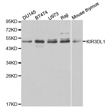
Figure 1. Western blot analysis of KIR3DL1 using anti-KIR3DL1 antibody (A02187-2). Electrophoresis was performed on a 5-20% SDS-PAGE gel at 70V (Stacking gel) / 90V (Resolving gel) for 2-3 hours. The sample well of each lane was loaded with 30 ug of sample under reducing conditions. Lane 1: human 293T whole cell lysates. After electrophoresis, proteins were transferred to a nitrocellulose membrane at 150 mA for 50-90 minutes. Blocked the membrane with 5% non-fat milk/TBS for 1.5 hour at RT. The membrane was incubated with rabbit anti-KIR3DL1 antigen affinity purified polyclonal antibody (Catalog # A02187-2) at 0.5 microg/mL overnight at 4°C, then washed with TBS-0.1%Tween 3 times with 5 minutes each and probed with a goat anti-rabbit IgG-HRP secondary antibody at a dilution of 1:5000 for 1.5 hour at RT. The signal is developed using an Enhanced Chemiluminescent detection (ECL) kit (Catalog # EK1002) with Tanon 5200 system. A specific band was detected for KIR3DL1 at approximately 49 kDa. The expected band size for KIR3DL1 is at 49 kDa.
Figure 1. Western blot analysis of KIR3DL1 using anti-KIR3DL1 antibody (A02187-2). Electrophoresis was performed on a 5-20% SDS-PAGE gel at 70V (Stacking gel) / 90V (Resolving gel) for 2-3 hours. The sample well of each lane was loaded with 30 ug of sample under reducing conditions. Lane 1: human 293T whole cell lysates. After electrophoresis, proteins were transferred to a nitrocellulose membrane at 150 mA for 50-90 minutes. Blocked the membrane with 5% non-fat milk/TBS for 1.5 hour at RT. The membrane was incubated with rabbit anti-KIR3DL1 antigen affinity purified polyclonal antibody (Catalog # A02187-2) at 0.5 microg/mL overnight at 4°C, then washed with TBS-0.1%Tween 3 times with 5 minutes each and probed with a goat anti-rabbit IgG-HRP secondary antibody at a dilution of 1:5000 for 1.5 hour at RT. The signal is developed using an Enhanced Chemiluminescent detection (ECL) kit (Catalog # EK1002) with Tanon 5200 system. A specific band was detected for KIR3DL1 at approximately 49 kDa. The expected band size for KIR3DL1 is at 49 kDa.
Anti-KIR3DL1 Antibody Picoband(r)
A02187-2-CARRIER-FREE
ApplicationsWestern Blot, ELISA
Product group Antibodies
ReactivityHuman
TargetKIR3DL1
Overview
- SupplierBoster Bio
- Product NameAnti-KIR3DL1 Antibody Picoband(r)
- Delivery Days Customer9
- ApplicationsWestern Blot, ELISA
- CertificationResearch Use Only
- ClonalityPolyclonal
- Concentration500 ug/ml
- Gene ID3811
- Target nameKIR3DL1
- Target descriptionkiller cell immunoglobulin like receptor, three Ig domains and long cytoplasmic tail 1
- Target synonymsCD158E1, KIR, KIR2DL5B, KIR3DL1/S1, NKAT-3, NKAT3, NKB1, NKB1B, killer cell immunoglobulin-like receptor 3DL1, CD158 antigen-like family member E, HLA-BW4-specific inhibitory NK cell receptor, KIR antigen 3DL1, killer cell immunoglobulin-like receptor, three domains, long cytoplasmic tail, 1, natural killer-associated transcript 3, p70 NK receptor CL-2/CL-11, p70 killer cell inhibitory receptor, p70 natural killer cell receptor clones CL-2/CL-11
- HostRabbit
- IsotypeIgG
- Protein IDP43629
- Protein NameKiller cell immunoglobulin-like receptor 3DL1
- Scientific DescriptionBoster Bio Anti-KIR3DL1 Antibody Picoband® catalog # A02187-2. Tested in ELISA, WB applications. This antibody reacts with Human. The brand Picoband indicates this is a premium antibody that guarantees superior quality, high affinity, and strong signals with minimal background in Western blot applications. Only our best-performing antibodies are designated as Picoband, ensuring unmatched performance.
- ReactivityHuman
- Storage Instruction-20°C,2°C to 8°C
- UNSPSC12352203
Related products
Product group Antibodies
Anti-KIR3DL1 AntibodyA30488
ApplicationsWestern Blot, ImmunoHistoChemistry
ReactivityHuman, Mouse
- SizePrice
Product group Antibodies
Anti-KIR3DL1 Antibody144-01617
ApplicationsWestern Blot
ReactivityHuman, Mouse
TargetKIR3DL1
- SizePrice
Product group Antibodies

KIR3DL1 AntibodyLS-C830797
ApplicationsELISA, ImmunoHistoChemistry
ReactivityHuman
TargetKIR3DL1
- SizePrice
Product group Antibodies
KIR3DL1 Polyclonal AntibodyBS-2421R
ApplicationsWestern Blot, ImmunoHistoChemistry, ImmunoHistoChemistry Frozen, ImmunoHistoChemistry Paraffin
ReactivityHuman, Mouse, Rat
- SizePrice
Product group Antibodies
KIR3DL1 AntibodyCSB-PA012364LA01HU
ApplicationsImmunoFluorescence, Western Blot, ELISA, ImmunoHistoChemistry
ReactivityHuman
TargetKIR3DL1
- SizePrice
Product group Antibodies
KIR3DL1 Polyclonal AntibodyCAC14854
ApplicationsImmunoFluorescence, Western Blot, ELISA, ImmunoHistoChemistry
TargetKIR3DL1
- SizePrice
Product group Antibodies
KIR3DL1 antibodyGTX106239
ApplicationsWestern Blot
ReactivityHuman
TargetKIR3DL1
- SizePrice