


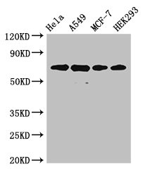
Figure 1. Western blot analysis of Ku70/XRCC6 using anti-Ku70/XRCC6 antibody (PB9520). Electrophoresis was performed on a 5-20% SDS-PAGE gel at 70V (Stacking gel) / 90V (Resolving gel) for 2-3 hours. The sample well of each lane was loaded with 30 ug of sample under reducing conditions. Lane 1: human LNCAP whole cell lysates, Lane 2: human Hela whole cell lysates, Lane 3: human 293T whole cell lysates, Lane 4: human HepG2 whole cell lysates, Lane 5: human Jurkat whole cell lysates, Lane 6: human K562 whole cell lysates, Lane 7: human A549 whole cell lysates, Lane 8: human A431 whole cell lysates. After electrophoresis, proteins were transferred to a nitrocellulose membrane at 150 mA for 50-90 minutes. Blocked the membrane with 5% non-fat milk/TBS for 1.5 hour at RT. The membrane was incubated with rabbit anti-Ku70/XRCC6 antigen affinity purified polyclonal antibody (Catalog # PB9520) at 0.5 microg/mL overnight at 4°C, then washed with TBS-0.1%Tween 3 times with 5 minutes each and probed with a goat anti-rabbit IgG-HRP secondary antibody at a dilution of 1:5000 for 1.5 hour at RT. The signal is developed using an Enhanced Chemiluminescent detection (ECL) kit (Catalog # EK1002) with Tanon 5200 system. A specific band was detected for Ku70/XRCC6 at approximately 70 kDa. The expected band size for Ku70/XRCC6 is at 70 kDa.
. Ku70/XRCC6 was detected in a paraffin-embedded section of human bladder cancer tissue. Heat mediated antigen retrieval was performed in EDTA buffer (pH 8.0, epitope retrieval solution). The tissue section was blocked with 10% goat serum. The tissue section was then incubated with 2 microg/ml rabbit anti-Ku70/XRCC6 Antibody (PB9520) overnight at 4°C. Peroxidase Conjugated Goat Anti-rabbit IgG was used as secondary antibody and incubated for 30 minutes at 37°C. The tissue section was developed using HRP Conjugated Rabbit IgG Super Vision Assay Kit (Catalog # SV0002) with DAB as the chromogen.")
. Ku70/XRCC6 was detected in a paraffin-embedded section of human bladder cancer tissue. Heat mediated antigen retrieval was performed in EDTA buffer (pH 8.0, epitope retrieval solution). The tissue section was blocked with 10% goat serum. The tissue section was then incubated with 2 microg/ml rabbit anti-Ku70/XRCC6 Antibody (PB9520) overnight at 4°C. Peroxidase Conjugated Goat Anti-rabbit IgG was used as secondary antibody and incubated for 30 minutes at 37°C. The tissue section was developed using HRP Conjugated Rabbit IgG Super Vision Assay Kit (Catalog # SV0002) with DAB as the chromogen.")
. Ku70/XRCC6 was detected in a paraffin-embedded section of human colon adenocarcinoma tissue. Heat mediated antigen retrieval was performed in EDTA buffer (pH 8.0, epitope retrieval solution). The tissue section was blocked with 10% goat serum. The tissue section was then incubated with 2 microg/ml rabbit anti-Ku70/XRCC6 Antibody (PB9520) overnight at 4°C. Peroxidase Conjugated Goat Anti-rabbit IgG was used as secondary antibody and incubated for 30 minutes at 37°C. The tissue section was developed using HRP Conjugated Rabbit IgG Super Vision Assay Kit (Catalog # SV0002) with DAB as the chromogen.")
. Ku70/XRCC6 was detected in a paraffin-embedded section of human colon adenocarcinoma tissue. Heat mediated antigen retrieval was performed in EDTA buffer (pH 8.0, epitope retrieval solution). The tissue section was blocked with 10% goat serum. The tissue section was then incubated with 2 microg/ml rabbit anti-Ku70/XRCC6 Antibody (PB9520) overnight at 4°C. Peroxidase Conjugated Goat Anti-rabbit IgG was used as secondary antibody and incubated for 30 minutes at 37°C. The tissue section was developed using HRP Conjugated Rabbit IgG Super Vision Assay Kit (Catalog # SV0002) with DAB as the chromogen.")
. Ku70/XRCC6 was detected in a paraffin-embedded section of human glioblastoma tissue. Heat mediated antigen retrieval was performed in EDTA buffer (pH 8.0, epitope retrieval solution). The tissue section was blocked with 10% goat serum. The tissue section was then incubated with 2 microg/ml rabbit anti-Ku70/XRCC6 Antibody (PB9520) overnight at 4°C. Peroxidase Conjugated Goat Anti-rabbit IgG was used as secondary antibody and incubated for 30 minutes at 37°C. The tissue section was developed using HRP Conjugated Rabbit IgG Super Vision Assay Kit (Catalog # SV0002) with DAB as the chromogen.")
. Ku70/XRCC6 was detected in a paraffin-embedded section of human glioblastoma tissue. Heat mediated antigen retrieval was performed in EDTA buffer (pH 8.0, epitope retrieval solution). The tissue section was blocked with 10% goat serum. The tissue section was then incubated with 2 microg/ml rabbit anti-Ku70/XRCC6 Antibody (PB9520) overnight at 4°C. Peroxidase Conjugated Goat Anti-rabbit IgG was used as secondary antibody and incubated for 30 minutes at 37°C. The tissue section was developed using HRP Conjugated Rabbit IgG Super Vision Assay Kit (Catalog # SV0002) with DAB as the chromogen.")
. Ku70/XRCC6 was detected in a paraffin-embedded section of human liver cancer tissue. Heat mediated antigen retrieval was performed in EDTA buffer (pH 8.0, epitope retrieval solution). The tissue section was blocked with 10% goat serum. The tissue section was then incubated with 2 microg/ml rabbit anti-Ku70/XRCC6 Antibody (PB9520) overnight at 4°C. Peroxidase Conjugated Goat Anti-rabbit IgG was used as secondary antibody and incubated for 30 minutes at 37°C. The tissue section was developed using HRP Conjugated Rabbit IgG Super Vision Assay Kit (Catalog # SV0002) with DAB as the chromogen.")
. Ku70/XRCC6 was detected in a paraffin-embedded section of human liver cancer tissue. Heat mediated antigen retrieval was performed in EDTA buffer (pH 8.0, epitope retrieval solution). The tissue section was blocked with 10% goat serum. The tissue section was then incubated with 2 microg/ml rabbit anti-Ku70/XRCC6 Antibody (PB9520) overnight at 4°C. Peroxidase Conjugated Goat Anti-rabbit IgG was used as secondary antibody and incubated for 30 minutes at 37°C. The tissue section was developed using HRP Conjugated Rabbit IgG Super Vision Assay Kit (Catalog # SV0002) with DAB as the chromogen.")
. Ku70/XRCC6 was detected in a paraffin-embedded section of human lung adenocarcinoma tissue. Heat mediated antigen retrieval was performed in EDTA buffer (pH 8.0, epitope retrieval solution). The tissue section was blocked with 10% goat serum. The tissue section was then incubated with 2 microg/ml rabbit anti-Ku70/XRCC6 Antibody (PB9520) overnight at 4°C. Peroxidase Conjugated Goat Anti-rabbit IgG was used as secondary antibody and incubated for 30 minutes at 37°C. The tissue section was developed using HRP Conjugated Rabbit IgG Super Vision Assay Kit (Catalog # SV0002) with DAB as the chromogen.")
Figure 1. Western blot analysis of Ku70/XRCC6 using anti-Ku70/XRCC6 antibody (PB9520). Electrophoresis was performed on a 5-20% SDS-PAGE gel at 70V (Stacking gel) / 90V (Resolving gel) for 2-3 hours. The sample well of each lane was loaded with 30 ug of sample under reducing conditions. Lane 1: human LNCAP whole cell lysates, Lane 2: human Hela whole cell lysates, Lane 3: human 293T whole cell lysates, Lane 4: human HepG2 whole cell lysates, Lane 5: human Jurkat whole cell lysates, Lane 6: human K562 whole cell lysates, Lane 7: human A549 whole cell lysates, Lane 8: human A431 whole cell lysates. After electrophoresis, proteins were transferred to a nitrocellulose membrane at 150 mA for 50-90 minutes. Blocked the membrane with 5% non-fat milk/TBS for 1.5 hour at RT. The membrane was incubated with rabbit anti-Ku70/XRCC6 antigen affinity purified polyclonal antibody (Catalog # PB9520) at 0.5 microg/mL overnight at 4°C, then washed with TBS-0.1%Tween 3 times with 5 minutes each and probed with a goat anti-rabbit IgG-HRP secondary antibody at a dilution of 1:5000 for 1.5 hour at RT. The signal is developed using an Enhanced Chemiluminescent detection (ECL) kit (Catalog # EK1002) with Tanon 5200 system. A specific band was detected for Ku70/XRCC6 at approximately 70 kDa. The expected band size for Ku70/XRCC6 is at 70 kDa.
Anti-Ku70/XRCC6 Antibody Picoband(r)
PB9520-CARRIER-FREE
ApplicationsFlow Cytometry, ImmunoFluorescence, Western Blot, ImmunoCytoChemistry, ImmunoHistoChemistry
Product group Antibodies
ReactivityHamster, Human
TargetXRCC6
Overview
- SupplierBoster Bio
- Product NameAnti-Ku70/XRCC6 Antibody Picoband(r)
- Delivery Days Customer9
- Application Supplier NoteTested Species: In-house tested species with positive results. By Heat: Boiling the paraffin sections in 10mM citrate buffer, pH6.0, for 20mins is required for the staining of formalin/paraffin sections. Other applications have not been tested. Optimal dilutions should be determined by end users.
- ApplicationsFlow Cytometry, ImmunoFluorescence, Western Blot, ImmunoCytoChemistry, ImmunoHistoChemistry
- CertificationResearch Use Only
- ClonalityPolyclonal
- Concentration500 ug/ml
- Gene ID2547
- Target nameXRCC6
- Target descriptionX-ray repair cross complementing 6
- Target synonymsCTC75, CTCBF, G22P1, KU70, ML8, TLAA, X-ray repair cross-complementing protein 6, 5'-dRP lyase Ku70, 5'-deoxyribose-5-phosphate lyase Ku70, 70 kDa subunit of Ku antigen, ATP-dependent DNA helicase 2 subunit 1, ATP-dependent DNA helicase II, 70 kDa subunit, CTC box binding factor 75 kDa subunit, DNA repair protein XRCC6, Ku autoantigen p70 subunit, Ku autoantigen, 70kDa, X-ray repair complementing defective repair in Chinese hamster cells 6, lupus Ku autoantigen protein p70, thyroid autoantigen 70kD (Ku antigen), thyroid autoantigen 70kDa (Ku antigen), thyroid-lupus autoantigen p70
- HostRabbit
- IsotypeIgG
- Protein IDP12956
- Protein NameX-ray repair cross-complementing protein 6
- Scientific DescriptionBoster Bio Anti-Ku70/XRCC6 Antibody Picoband® catalog # PB9520. Tested in Flow Cytometry, IF, IHC, ICC, WB applications. This antibody reacts with Human. The brand Picoband indicates this is a premium antibody that guarantees superior quality, high affinity, and strong signals with minimal background in Western blot applications. Only our best-performing antibodies are designated as Picoband, ensuring unmatched performance.
- ReactivityHamster, Human
- Storage Instruction-20°C,2°C to 8°C
- UNSPSC12352203
Related products
Product group Antibodies
XRCC6 AntibodyCSB-PA01617A0RB
ApplicationsImmunoFluorescence, Western Blot, ELISA, ImmunoHistoChemistry
ReactivityHuman
TargetXRCC6
- SizePrice
Product group Antibodies
Anti-XRCC6 Antibody144-00883
ApplicationsImmunoFluorescence, Western Blot, ImmunoHistoChemistry
ReactivityHuman, Monkey, Mouse, Rat
TargetXRCC6
- SizePrice
Product group Antibodies
Anti-Ku70 AntibodyA95361
ApplicationsImmunoFluorescence, Western Blot, ELISA, ImmunoHistoChemistry
ReactivityHuman, Mouse, Rat
- SizePrice
Product group Antibodies

Goat anti-KU70 / XRCC6EB07858
ApplicationsWestern Blot, ELISA
ReactivityHuman
TargetXRCC6
- SizePrice

Product group Antibodies
Anti-XRCC6 AntibodyHPA047549
ApplicationsWestern Blot, ImmunoCytoChemistry, ImmunoHistoChemistry
ReactivityHuman
TargetXRCC6
- SizePrice
Product group Antibodies

XRCC6 / Ku70 Antibody (C-Terminus)LS-C358896
ApplicationsWestern Blot
ReactivityHuman, Monkey
TargetXRCC6
- SizePrice
Product group Antibodies
XRCC6 Polyclonal AntibodyCAC12912
ApplicationsImmunoFluorescence, Western Blot, ELISA, ImmunoHistoChemistry
TargetXRCC6
- SizePrice
Product group Antibodies
Ku70 antibodyGTX101820
ApplicationsImmunoFluorescence, ImmunoPrecipitation, Western Blot, ImmunoCytoChemistry, ImmunoHistoChemistry, ImmunoHistoChemistry Paraffin, Other Application
ReactivityHuman, Mouse
TargetXRCC6
- SizePrice