


Figure 1. IHC analysis of L1CAM using anti-L1CAM antibody (A00729-1). L1CAM was detected in paraffin-embedded section of mouse brain tissue. Heat mediated antigen retrieval was performed in citrate buffer (pH6, epitope retrieval solution) for 20 mins. The tissue section was blocked with 10% goat serum. The tissue section was then incubated with 1microg/ml rabbit anti-L1CAM Antibody (A00729-1) overnight at 4°C. Biotinylated goat anti-rabbit IgG was used as secondary antibody and incubated for 30 minutes at 37°C. The tissue section was developed using Strepavidin-Biotin-Complex (SABC)(Catalog # SA1022) with DAB as the chromogen.
. L1CAM was detected in paraffin-embedded section of mouse brain tissue. Heat mediated antigen retrieval was performed in citrate buffer (pH6, epitope retrieval solution) for 20 mins. The tissue section was blocked with 10% goat serum. The tissue section was then incubated with 1microg/ml rabbit anti-L1CAM Antibody (A00729-1) overnight at 4°C. Biotinylated goat anti-rabbit IgG was used as secondary antibody and incubated for 30 minutes at 37°C. The tissue section was developed using Strepavidin-Biotin-Complex (SABC)(Catalog # SA1022) with DAB as the chromogen.")
. L1CAM was detected in paraffin-embedded section of rat brain tissue. Heat mediated antigen retrieval was performed in citrate buffer (pH6, epitope retrieval solution) for 20 mins. The tissue section was blocked with 10% goat serum. The tissue section was then incubated with 1microg/ml rabbit anti-L1CAM Antibody (A00729-1) overnight at 4°C. Biotinylated goat anti-rabbit IgG was used as secondary antibody and incubated for 30 minutes at 37°C. The tissue section was developed using Strepavidin-Biotin-Complex (SABC)(Catalog # SA1022) with DAB as the chromogen.")
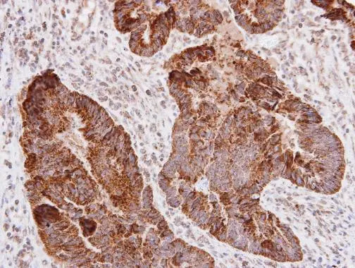
. L1CAM was detected in paraffin-embedded section of human colon cancer tissue. Heat mediated antigen retrieval was performed in citrate buffer (pH6, epitope retrieval solution) for 20 mins. The tissue section was blocked with 10% goat serum. The tissue section was then incubated with 1microg/ml rabbit anti-L1CAM Antibody (A00729-1) overnight at 4°C. Biotinylated goat anti-rabbit IgG was used as secondary antibody and incubated for 30 minutes at 37°C. The tissue section was developed using Strepavidin-Biotin-Complex (SABC)(Catalog # SA1022) with DAB as the chromogen.")
. L1CAM was detected in paraffin-embedded section of human glioma tissue. Heat mediated antigen retrieval was performed in citrate buffer (pH6, epitope retrieval solution) for 20 mins. The tissue section was blocked with 10% goat serum. The tissue section was then incubated with 1microg/ml rabbit anti-L1CAM Antibody (A00729-1) overnight at 4°C. Biotinylated goat anti-rabbit IgG was used as secondary antibody and incubated for 30 minutes at 37°C. The tissue section was developed using Strepavidin-Biotin-Complex (SABC)(Catalog # SA1022) with DAB as the chromogen.")
. L1CAM was detected in paraffin-embedded section of human lung cancer tissue. Heat mediated antigen retrieval was performed in citrate buffer (pH6, epitope retrieval solution) for 20 mins. The tissue section was blocked with 10% goat serum. The tissue section was then incubated with 1microg/ml rabbit anti-L1CAM Antibody (A00729-1) overnight at 4°C. Biotinylated goat anti-rabbit IgG was used as secondary antibody and incubated for 30 minutes at 37°C. The tissue section was developed using Strepavidin-Biotin-Complex (SABC)(Catalog # SA1022) with DAB as the chromogen.")
. L1CAM was detected in paraffin-embedded section of human mammary cancer tissue. Heat mediated antigen retrieval was performed in citrate buffer (pH6, epitope retrieval solution) for 20 mins. The tissue section was blocked with 10% goat serum. The tissue section was then incubated with 1microg/ml rabbit anti-L1CAM Antibody (A00729-1) overnight at 4°C. Biotinylated goat anti-rabbit IgG was used as secondary antibody and incubated for 30 minutes at 37°C. The tissue section was developed using Strepavidin-Biotin-Complex (SABC)(Catalog # SA1022) with DAB as the chromogen.")
Figure 1. IHC analysis of L1CAM using anti-L1CAM antibody (A00729-1). L1CAM was detected in paraffin-embedded section of mouse brain tissue. Heat mediated antigen retrieval was performed in citrate buffer (pH6, epitope retrieval solution) for 20 mins. The tissue section was blocked with 10% goat serum. The tissue section was then incubated with 1microg/ml rabbit anti-L1CAM Antibody (A00729-1) overnight at 4°C. Biotinylated goat anti-rabbit IgG was used as secondary antibody and incubated for 30 minutes at 37°C. The tissue section was developed using Strepavidin-Biotin-Complex (SABC)(Catalog # SA1022) with DAB as the chromogen.
Anti-L1CAM Antibody
A00729-1-CARRIER-FREE
ApplicationsImmunoHistoChemistry
Product group Antibodies
ReactivityHuman, Mouse, Rat
TargetL1CAM
Overview
- SupplierBoster Bio
- Product NameAnti-L1CAM Antibody
- Delivery Days Customer9
- ApplicationsImmunoHistoChemistry
- CertificationResearch Use Only
- ClonalityPolyclonal
- Concentration500 ug/ml
- Gene ID3897
- Target nameL1CAM
- Target descriptionL1 cell adhesion molecule
- Target synonymsCAML1, CD171, HSAS, HSAS1, HYCX, MASA, MIC5, N-CAM-L1, N-CAML1, NCAM-L1, S10, SPG1, neural cell adhesion molecule L1, antigen identified by monoclonal antibody R1
- HostRabbit
- IsotypeIgG
- Protein IDP32004
- Protein NameNeural cell adhesion molecule L1
- Scientific DescriptionBoster Bio Anti-L1CAM Antibody Picoband® catalog # A00729-1. Tested in IHC applications. This antibody reacts with Human, Mouse, Rat.
- ReactivityHuman, Mouse, Rat
- Storage Instruction-20°C,2°C to 8°C
- UNSPSC12352203
Related products
Product group Antibodies
Anti-L1CAM Antibody144-08555
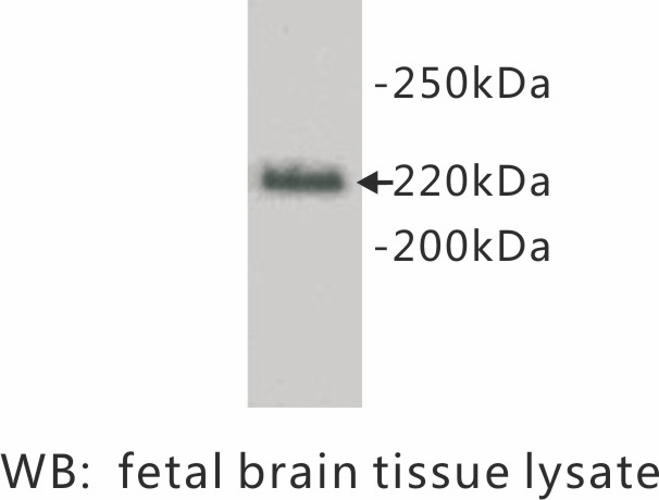
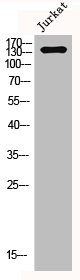
ApplicationsWestern Blot
ReactivityHuman, Mouse, Rat
TargetL1CAM
- SizePrice
Product group Antibodies

Anti-CD171 [L1-14.10]Ab01427-1.1
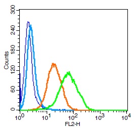
ApplicationsFlow Cytometry, ImmunoFluorescence, ImmunoPrecipitation, Western Blot, ImmunoHistoChemistry
ReactivityHuman
TargetL1CAM
- SizePrice

Product group Antibodies
Anti-L1CAM AntibodyAMAB91829
ApplicationsImmunoHistoChemistry
ReactivityHuman
TargetL1CAM
- SizePrice
Product group Antibodies
ApplicationsWestern Blot, ImmunoCytoChemistry
ReactivityHuman
- SizePrice
Product group Antibodies

L1CAM Antibody (clone 014, FITC)LS-C818857
ApplicationsFlow Cytometry
ReactivityHuman
TargetL1CAM
- SizePrice
Product group Antibodies
CD171/L1CAM Polyclonal AntibodyBS-1996R
ApplicationsFlow Cytometry, ImmunoFluorescence, ELISA, ImmunoCytoChemistry, ImmunoHistoChemistry, ImmunoHistoChemistry Frozen, ImmunoHistoChemistry Paraffin
ReactivityBovine, Canine, Chicken, Equine, Human, Mouse, Porcine, Rabbit, Rat
TargetL1CAM
- SizePrice
Product group Antibodies
L1CAM AntibodyCSB-PA003380
ApplicationsImmunoFluorescence, Western Blot, ELISA, ImmunoHistoChemistry
ReactivityHuman, Mouse, Rat
TargetL1CAM
- SizePrice
Product group Antibodies
L1Cam Polyclonal AntibodyCAC10396
ApplicationsImmunoFluorescence, Western Blot, ELISA, ImmunoHistoChemistry
ReactivityMouse
TargetL1CAM
- SizePrice
Product group Antibodies
L1CAM antibodyGTX129010
ApplicationsWestern Blot, ImmunoHistoChemistry, ImmunoHistoChemistry Paraffin
ReactivityHuman
TargetL1CAM
- SizePrice