

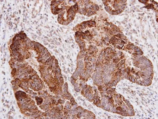
L1CAM antibody detects L1CAM protein at cell membrane on human colon carcinoma by immunohistochemical analysis. Sample: Paraffin-embedded human colon carcinoma. L1CAM antibody (GTX129010) diluted at 1:250.
Antigen Retrieval: Trilogy? (EDTA based, pH 8.0) buffer, 15min
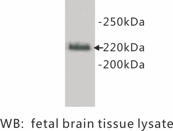
 were separated by 5% SDS-PAGE, and the membrane was blotted with L1CAM antibody (GTX129010) diluted at 1:1000. The HRP-conjugated anti-rabbit IgG antibody (GTX213110-01) was used to detect the primary antibody. Corresponding RNA expression data for the same cell lines are based on Human Protein Atlas program.")
L1CAM antibody detects L1CAM protein at cell membrane on human colon carcinoma by immunohistochemical analysis. Sample: Paraffin-embedded human colon carcinoma. L1CAM antibody (GTX129010) diluted at 1:250.
Antigen Retrieval: Trilogy? (EDTA based, pH 8.0) buffer, 15min
L1CAM antibody
GTX129010
ApplicationsWestern Blot, ImmunoHistoChemistry, ImmunoHistoChemistry Paraffin
Product group Antibodies
ReactivityHuman
TargetL1CAM
Overview
- SupplierGeneTex
- Product NameL1CAM antibody
- Delivery Days Customer9
- Application Supplier NoteWB: 1:500-1:3000. IHC-P: 1:100-1:1000. *Optimal dilutions/concentrations should be determined by the researcher.Not tested in other applications.
- ApplicationsWestern Blot, ImmunoHistoChemistry, ImmunoHistoChemistry Paraffin
- CertificationResearch Use Only
- ClonalityPolyclonal
- Concentration1 mg/ml
- ConjugateUnconjugated
- Gene ID3897
- Target nameL1CAM
- Target descriptionL1 cell adhesion molecule
- Target synonymsCAML1, CD171, HSAS, HSAS1, HYCX, MASA, MIC5, N-CAM-L1, N-CAML1, NCAM-L1, S10, SPG1, neural cell adhesion molecule L1, antigen identified by monoclonal antibody R1
- HostRabbit
- IsotypeIgG
- Protein IDP32004
- Protein NameNeural cell adhesion molecule L1
- Scientific DescriptionThe protein encoded by this gene is an axonal glycoprotein belonging to the immunoglobulin supergene family. The ectodomain, consisting of several immunoglobulin-like domains and fibronectin-like repeats (type III), is linked via a single transmembrane sequence to a conserved cytoplasmic domain. This cell adhesion molecule plays an important role in nervous system development, including neuronal migration and differentiation. Mutations in the gene cause X-linked neurological syndromes known as CRASH (corpus callosum hypoplasia, retardation, aphasia, spastic paraplegia and hydrocephalus). Alternative splicing of this gene results in multiple transcript variants, some of which include an alternate exon that is considered to be specific to neurons. [provided by RefSeq, May 2013]
- ReactivityHuman
- Storage Instruction-20°C or -80°C,2°C to 8°C
- UNSPSC41116161
Datasheet
Related products

Product group Antibodies
Anti-L1CAM AntibodyA00729-1-CARRIER-FREE
ApplicationsImmunoHistoChemistry
ReactivityHuman, Mouse, Rat
TargetL1CAM
- SizePrice
Product group Antibodies
Anti-L1CAM Antibody144-08555
ApplicationsWestern Blot
ReactivityHuman, Mouse, Rat
TargetL1CAM
- SizePrice
Product group Antibodies

Anti-CD171 [L1-14.10]Ab01427-1.1
ApplicationsFlow Cytometry, ImmunoFluorescence, ImmunoPrecipitation, Western Blot, ImmunoHistoChemistry
ReactivityHuman
TargetL1CAM
- SizePrice

Product group Antibodies
Anti-L1CAM AntibodyAMAB91829
ApplicationsImmunoHistoChemistry
ReactivityHuman
TargetL1CAM
- SizePrice
Product group Antibodies
ApplicationsWestern Blot, ImmunoCytoChemistry
ReactivityHuman
- SizePrice
Product group Antibodies

L1CAM Antibody (clone 014, FITC)LS-C818857
ApplicationsFlow Cytometry
ReactivityHuman
TargetL1CAM
- SizePrice
Product group Antibodies
CD171/L1CAM Polyclonal AntibodyBS-1996R
ApplicationsFlow Cytometry, ImmunoFluorescence, ELISA, ImmunoCytoChemistry, ImmunoHistoChemistry, ImmunoHistoChemistry Frozen, ImmunoHistoChemistry Paraffin
ReactivityBovine, Canine, Chicken, Equine, Human, Mouse, Porcine, Rabbit, Rat
TargetL1CAM
- SizePrice

Product group Antibodies
L1CAM AntibodyCSB-PA003380
ApplicationsImmunoFluorescence, Western Blot, ELISA, ImmunoHistoChemistry
ReactivityHuman, Mouse, Rat
TargetL1CAM
- SizePrice
Product group Antibodies
L1Cam Polyclonal AntibodyCAC10396
ApplicationsImmunoFluorescence, Western Blot, ELISA, ImmunoHistoChemistry
ReactivityMouse
TargetL1CAM
- SizePrice