

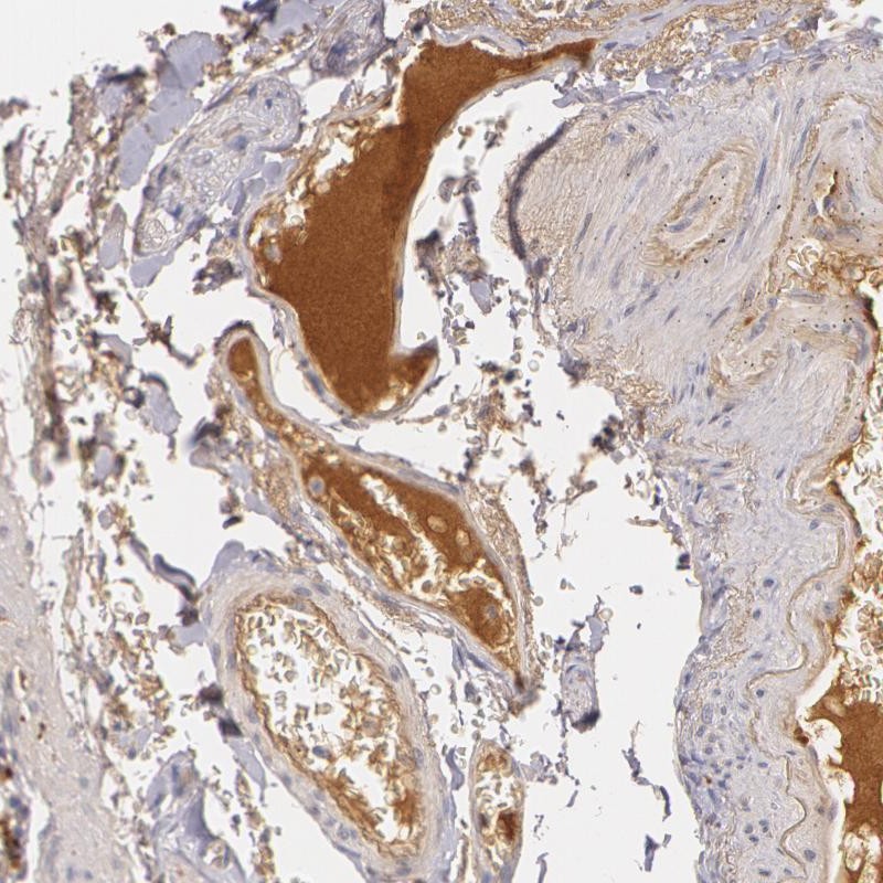
Immunohistochemical staining of human small intestine shows positivity in plasma.
Immunohistochemical staining of human small intestine shows positivity in plasma.
Anti-LBP Antibody
HPA001508
ApplicationsImmunoHistoChemistry
Product group Antibodies
ReactivityHuman
TargetLBP
Overview
- SupplierAtlas Antibodies
- Product NameAnti-LBP Antibody
- Delivery Days Customer4
- ApplicationsImmunoHistoChemistry
- CertificationResearch Use Only
- ClonalityPolyclonal
- ConjugateUnconjugated
- Gene ID3929
- Target nameLBP
- Target descriptionlipopolysaccharide binding protein
- Target synonymsBPIFD2, lipopolysaccharide-binding protein, BPI fold containing family D, member 2, LPS-binding protein
- HostRabbit
- IsotypeIgG
- Protein IDP18428
- Protein NameLipopolysaccharide-binding protein
- Scientific DescriptionRecombinant Protein Epitope Signature Tag (PrEST) antigen sequence
- ReactivityHuman
- Storage Instruction-20°C,2°C to 8°C
- UNSPSC41116161
Datasheet
MSDS
Related products
Product group Antibodies
Anti-LBP AntibodyA98491
ApplicationsWestern Blot, ELISA
ReactivityHuman, Mouse, Rat
- SizePrice

Product group Antibodies
Anti-LBP Antibody Picoband(r)A00809-1-CARRIER-FREE
ApplicationsWestern Blot
ReactivityHuman, Mouse, Rat
TargetLBP
- SizePrice
Product group Antibodies
Anti-LBP Antibody144-64959
ApplicationsWestern Blot, ImmunoHistoChemistry
ReactivityHuman, Mouse, Rat
TargetLBP
- SizePrice
Product group Antibodies
LBP AntibodyCSB-PA009787
ApplicationsWestern Blot, ELISA
ReactivityHuman, Mouse, Rat
TargetLBP
- SizePrice
Product group Antibodies
ApplicationsImmunoPrecipitation, Western Blot, ImmunoCytoChemistry, ImmunoHistoChemistry
ReactivityRat
TargetLBP
- SizePrice

Product group Antibodies
LBP antibody [N3C3]GTX105154
ApplicationsWestern Blot, ImmunoHistoChemistry, ImmunoHistoChemistry Paraffin
ReactivityHuman
TargetLBP
- SizePrice
Product group Antibodies
Anti-LBP AntibodyCAB17507
ApplicationsImmunoFluorescence, Western Blot, ELISA, ImmunoCytoChemistry
ReactivityHuman
TargetLBP
- SizePrice