


Figure 1. Western blot analysis of LCMT2 using anti-LCMT2 antibody (A14797-1). Electrophoresis was performed on a 5-20% SDS-PAGE gel at 70V (Stacking gel) / 90V (Resolving gel) for 2-3 hours. The sample well of each lane was loaded with 30 ug of sample under reducing conditions. Lane 1: human Hacat whole cell lysates, Lane 2: human Hela whole cell lysates, Lane 3: human THP-1 whole cell lysates, Lane 4: human SH-SY5Y whole cell lysates, Lane 5: rat brain tissue lysates, Lane 6: mouse brain tissue lysates. After electrophoresis, proteins were transferred to a nitrocellulose membrane at 150 mA for 50-90 minutes. Blocked the membrane with 5% non-fat milk/TBS for 1.5 hour at RT. The membrane was incubated with rabbit anti-LCMT2 antigen affinity purified polyclonal antibody (Catalog # A14797-1) at 0.25 microg/mL overnight at 4°C, then washed with TBS-0.1%Tween 3 times with 5 minutes each and probed with a goat anti-rabbit IgG-HRP secondary antibody at a dilution of 1:5000 for 1.5 hour at RT. The signal is developed using an Enhanced Chemiluminescent detection (ECL) kit (Catalog # EK1002) with Tanon 5200 system. A specific band was detected for LCMT2 at approximately 76 kDa. The expected band size for LCMT2 is at 76 kDa.
. LCMT2 was detected in an immunocytochemical section of U2OS cells. Enzyme antigen retrieval was performed using IHC enzyme antigen retrieval reagent (AR0022) for 15 mins. The cells were blocked with 10% goat serum. And then incubated with 5 microg/mL rabbit anti-LCMT2 Antibody (A14797-1) overnight at 4°C. DyLight®488 Conjugated Goat Anti-Rabbit IgG (BA1127) was used as secondary antibody at 1:500 dilution and incubated for 30 minutes at 37°C. The section was counterstained with DAPI. Visualize using a fluorescence microscope and filter sets appropriate for the label used.")
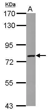
Figure 1. Western blot analysis of LCMT2 using anti-LCMT2 antibody (A14797-1). Electrophoresis was performed on a 5-20% SDS-PAGE gel at 70V (Stacking gel) / 90V (Resolving gel) for 2-3 hours. The sample well of each lane was loaded with 30 ug of sample under reducing conditions. Lane 1: human Hacat whole cell lysates, Lane 2: human Hela whole cell lysates, Lane 3: human THP-1 whole cell lysates, Lane 4: human SH-SY5Y whole cell lysates, Lane 5: rat brain tissue lysates, Lane 6: mouse brain tissue lysates. After electrophoresis, proteins were transferred to a nitrocellulose membrane at 150 mA for 50-90 minutes. Blocked the membrane with 5% non-fat milk/TBS for 1.5 hour at RT. The membrane was incubated with rabbit anti-LCMT2 antigen affinity purified polyclonal antibody (Catalog # A14797-1) at 0.25 microg/mL overnight at 4°C, then washed with TBS-0.1%Tween 3 times with 5 minutes each and probed with a goat anti-rabbit IgG-HRP secondary antibody at a dilution of 1:5000 for 1.5 hour at RT. The signal is developed using an Enhanced Chemiluminescent detection (ECL) kit (Catalog # EK1002) with Tanon 5200 system. A specific band was detected for LCMT2 at approximately 76 kDa. The expected band size for LCMT2 is at 76 kDa.
Anti-LCMT2 Antibody Picoband(r)
A14797-1-CARRIER-FREE
ApplicationsImmunoFluorescence, Western Blot, ELISA, ImmunoCytoChemistry
Product group Antibodies
ReactivityHuman, Mouse, Rat
TargetLCMT2
Overview
- SupplierBoster Bio
- Product NameAnti-LCMT2 Antibody Picoband(r)
- Delivery Days Customer9
- ApplicationsImmunoFluorescence, Western Blot, ELISA, ImmunoCytoChemistry
- CertificationResearch Use Only
- ClonalityPolyclonal
- Concentration500 ug/ml
- Gene ID9836
- Target nameLCMT2
- Target descriptionleucine carboxyl methyltransferase 2
- Target synonymsPPM2, TYW4, tRNA wybutosine-synthesizing protein 4, p21WAF1/CIP1 promoter-interacting protein, tRNA(Phe) (7-(3-amino-3-(methoxycarbonyl)propyl)wyosine(37)-N)-methoxycarbonyltransferase, tRNA(Phe) (7-(3-amino-3-carboxypropyl)wyosine(37)-O)-methyltransferase, tRNA-Wybutosine-synthesizing protein 4, tRNA-yW synthesizing protein 4
- HostRabbit
- IsotypeIgG
- Protein IDO60294
- Protein NametRNA wybutosine-synthesizing protein 4
- Scientific DescriptionBoster Bio Anti-LCMT2 Antibody Picoband® catalog # A14797-1. Tested in ELISA, IF, ICC, WB applications. This antibody reacts with Human, Mouse, Rat. The brand Picoband indicates this is a premium antibody that guarantees superior quality, high affinity, and strong signals with minimal background in Western blot applications. Only our best-performing antibodies are designated as Picoband, ensuring unmatched performance.
- ReactivityHuman, Mouse, Rat
- Storage Instruction-20°C,2°C to 8°C
- UNSPSC12352203
Related products
Product group Antibodies

LCMT2 / YW4 AntibodyLS-C830536
ApplicationsELISA, ImmunoHistoChemistry
ReactivityHuman, Mouse, Rat
TargetLCMT2
- SizePrice

Product group Antibodies
Anti-LCMT2 AntibodyHPA039401
ApplicationsImmunoHistoChemistry
ReactivityHuman
TargetLCMT2
- SizePrice
Product group Antibodies
LCMT2 antibodyGTX122895
ApplicationsWestern Blot, ImmunoHistoChemistry, ImmunoHistoChemistry Paraffin
ReactivityHuman, Mouse
TargetLCMT2
- SizePrice
Product group Antibodies
Anti-p21WAF1 (Center H450) Antibody102-20929
ApplicationsWestern Blot
TargetLCMT2
- SizePrice