


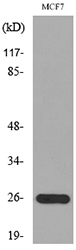
Figure 1. Western blot analysis of Galectin-3/LGALS3 using anti-Galectin-3/LGALS3 antibody (A00621-1). Electrophoresis was performed on a 5-20% SDS-PAGE gel at 70V (Stacking gel) / 90V (Resolving gel) for 2-3 hours. The sample well of each lane was loaded with 30 ug of sample under reducing conditions. Lane 1: human Hela whole cell lysates, Lane 2: human A431 whole cell lysates, Lane 3: human A549 whole cell lysates, Lane 4: human MCF-7 whole cell lysates, Lane 5: rat stomach tissue lysates, Lane 6: mouse stomach tissue lysates. After electrophoresis, proteins were transferred to a nitrocellulose membrane at 150 mA for 50-90 minutes. Blocked the membrane with 5% non-fat milk/TBS for 1.5 hour at RT. The membrane was incubated with rabbit anti-Galectin-3/LGALS3 antigen affinity purified polyclonal antibody (Catalog # A00621-1) at 0.5 microg/mL overnight at 4°C, then washed with TBS-0.1%Tween 3 times with 5 minutes each and probed with a goat anti-rabbit IgG-HRP secondary antibody at a dilution of 1:5000 for 1.5 hour at RT. The signal is developed using an Enhanced Chemiluminescent detection (ECL) kit (Catalog # EK1002) with Tanon 5200 system. A specific band was detected for Galectin-3/LGALS3 at approximately 29 kDa. The expected band size for Galectin-3/LGALS3 is at 26 kDa.
. Galectin-3/LGALS3 was detected in an immunocytochemical section of HELA cells. Enzyme antigen retrieval was performed using IHC enzyme antigen retrieval reagent (AR0022) for 15 mins. The cells were blocked with 10% goat serum. And then incubated with 5 microg/mL rabbit anti-Galectin-3/LGALS3 Antibody (A00621-1) overnight at 4°C. DyLight®488 Conjugated Goat Anti-Rabbit IgG (BA1127) was used as secondary antibody at 1:500 dilution and incubated for 30 minutes at 37°C. The section was counterstained with DAPI. Visualize using a fluorescence microscope and filter sets appropriate for the label used.")
Figure 1. Western blot analysis of Galectin-3/LGALS3 using anti-Galectin-3/LGALS3 antibody (A00621-1). Electrophoresis was performed on a 5-20% SDS-PAGE gel at 70V (Stacking gel) / 90V (Resolving gel) for 2-3 hours. The sample well of each lane was loaded with 30 ug of sample under reducing conditions. Lane 1: human Hela whole cell lysates, Lane 2: human A431 whole cell lysates, Lane 3: human A549 whole cell lysates, Lane 4: human MCF-7 whole cell lysates, Lane 5: rat stomach tissue lysates, Lane 6: mouse stomach tissue lysates. After electrophoresis, proteins were transferred to a nitrocellulose membrane at 150 mA for 50-90 minutes. Blocked the membrane with 5% non-fat milk/TBS for 1.5 hour at RT. The membrane was incubated with rabbit anti-Galectin-3/LGALS3 antigen affinity purified polyclonal antibody (Catalog # A00621-1) at 0.5 microg/mL overnight at 4°C, then washed with TBS-0.1%Tween 3 times with 5 minutes each and probed with a goat anti-rabbit IgG-HRP secondary antibody at a dilution of 1:5000 for 1.5 hour at RT. The signal is developed using an Enhanced Chemiluminescent detection (ECL) kit (Catalog # EK1002) with Tanon 5200 system. A specific band was detected for Galectin-3/LGALS3 at approximately 29 kDa. The expected band size for Galectin-3/LGALS3 is at 26 kDa.
Anti-LGALS3 Antibody Picoband(r)
A00621-1-CARRIER-FREE
ApplicationsImmunoFluorescence, Western Blot, ELISA, ImmunoCytoChemistry
Product group Antibodies
ReactivityHuman, Mouse, Rat
TargetLGALS3
Overview
- SupplierBoster Bio
- Product NameAnti-LGALS3 Antibody Picoband(r)
- Delivery Days Customer9
- ApplicationsImmunoFluorescence, Western Blot, ELISA, ImmunoCytoChemistry
- CertificationResearch Use Only
- ClonalityPolyclonal
- Concentration500 ug/ml
- Gene ID3958
- Target nameLGALS3
- Target descriptiongalectin 3
- Target synonymsCBP35, GAL3, GALBP, GALIG, L31, LGALS2, MAC2, galectin-3, 35 kDa lectin, IgE-binding protein, MAC-2 antigen, advanced glycation end-product receptor 3, carbohydrate-binding protein 35, epididymis secretory sperm binding protein, galactose-specific lectin 3, laminin-binding protein, lectin L-29, lectin, galactoside-binding, soluble, 3
- HostRabbit
- Protein IDP17931
- Protein NameGalectin-3
- Scientific DescriptionBoster Bio Anti-LGALS3 Antibody Picoband® catalog # A00621-1. Tested in WB, ICC/IF, ELISA applications. This antibody reacts with Human, Mouse, Rat. The brand Picoband indicates this is a premium antibody that guarantees superior quality, high affinity, and strong signals with minimal background in Western blot applications. Only our best-performing antibodies are designated as Picoband, ensuring unmatched performance.
- ReactivityHuman, Mouse, Rat
- Storage Instruction-20°C,2°C to 8°C
- UNSPSC12352203
Related products

Product group Antibodies
ApplicationsImmunoCytoChemistry
ReactivityHuman
TargetLGALS3
- SizePrice
Product group Antibodies
Anti-LGALS3 AntibodyA100515
ApplicationsWestern Blot, ELISA
ReactivityHuman
- SizePrice
Product group Antibodies
Anti-Galectin-3 Antibody130-10090
ApplicationsELISA
ReactivityHuman
TargetLGALS3
- SizePrice
Product group Antibodies
References
Galectin 3 Polyclonal AntibodyBS-0721R
ApplicationsFlow Cytometry, ImmunoFluorescence, ELISA, ImmunoCytoChemistry, ImmunoHistoChemistry, ImmunoHistoChemistry Frozen, ImmunoHistoChemistry Paraffin
ReactivityHuman, Mouse
TargetLGALS3
- SizePrice
Product group Antibodies
LGALS3 Monoclonal AntibodyCSB-MA000198
ApplicationsWestern Blot, ELISA, ImmunoHistoChemistry
ReactivityHuman
TargetLGALS3
- SizePrice
Product group Antibodies

Goat anti-Galectin 3EB07710
ApplicationsWestern Blot, ELISA
ReactivityHuman, Mouse, Rat
TargetLGALS3
- SizePrice
Product group Antibodies
LGALS3 Polyclonal AntibodyCAC13799
ApplicationsWestern Blot, ELISA, ImmunoHistoChemistry
TargetLGALS3
- SizePrice
Product group Antibodies

LGALS3 / Galectin 3 AntibodyLS-C343056
ApplicationsWestern Blot, ELISA
ReactivityHuman
TargetLGALS3
- SizePrice

Product group Antibodies
Anti-LGALS3-25ulHPA003162
ApplicationsWestern Blot, ImmunoHistoChemistry
ReactivityHuman
- SizePrice