


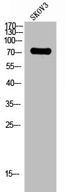
Figure 1. Western blot analysis of LILRB1 using anti-LILRB1 antibody (A03208-1). Electrophoresis was performed on a 5-20% SDS-PAGE gel at 70V (Stacking gel) / 90V (Resolving gel) for 2-3 hours. The sample well of each lane was loaded with 30 ug of sample under reducing conditions. Lane 1: human THP-1 whole cell lysates, Lane 2: human Raji whole cell lysates. After electrophoresis, proteins were transferred to a nitrocellulose membrane at 150 mA for 50-90 minutes. Blocked the membrane with 5% non-fat milk/TBS for 1.5 hour at RT. The membrane was incubated with rabbit anti-LILRB1 antigen affinity purified polyclonal antibody (Catalog # A03208-1) at 0.5 microg/mL overnight at 4°C, then washed with TBS-0.1%Tween 3 times with 5 minutes each and probed with a goat anti-rabbit IgG-HRP secondary antibody at a dilution of 1:5000 for 1.5 hour at RT. The signal is developed using an Enhanced Chemiluminescent detection (ECL) kit (Catalog # EK1002) with Tanon 5200 system. A specific band was detected for LILRB1 at approximately 71 kDa. The expected band size for LILRB1 is at 71 kDa.
. LILRB1 was detected in a paraffin-embedded section of human liver cancer tissue. Heat mediated antigen retrieval was performed in EDTA buffer (pH 8.0, epitope retrieval solution). The tissue section was blocked with 10% goat serum. The tissue section was then incubated with 2 microg/ml rabbit anti-LILRB1 Antibody (A03208-1) overnight at 4°C. Biotinylated goat anti-rabbit IgG was used as secondary antibody and incubated for 30 minutes at 37°C. The tissue section was developed using Strepavidin-Biotin-Complex (SABC) (Catalog # SA1022) with DAB as the chromogen.")
. LILRB1 was detected in a paraffin-embedded section of human lymphoma tissue. Heat mediated antigen retrieval was performed in EDTA buffer (pH 8.0, epitope retrieval solution). The tissue section was blocked with 10% goat serum. The tissue section was then incubated with 2 microg/ml rabbit anti-LILRB1 Antibody (A03208-1) overnight at 4°C. Biotinylated goat anti-rabbit IgG was used as secondary antibody and incubated for 30 minutes at 37°C. The tissue section was developed using Strepavidin-Biotin-Complex (SABC) (Catalog # SA1022) with DAB as the chromogen.")
. Overlay histogram showing HL-60 cells stained with A03208-1 (Blue line). The cells were fixed with 4% paraformaldehyde and blocked with 10% normal goat serum. And then incubated with rabbit anti-LILRB1 Antibody (A03208-1, 1 microg/1x106 cells) for 30 min at 20°C. DyLight®488 conjugated goat anti-rabbit IgG (BA1127, 5-10 microg/1x106 cells) was used as secondary antibody for 30 minutes at 20°C. Isotype control antibody (Green line) was rabbit IgG (1 microg/1x106) used under the same conditions. Unlabelled sample without incubation with primary antibody and secondary antibody (Red line) was used as a blank control.")
Figure 1. Western blot analysis of LILRB1 using anti-LILRB1 antibody (A03208-1). Electrophoresis was performed on a 5-20% SDS-PAGE gel at 70V (Stacking gel) / 90V (Resolving gel) for 2-3 hours. The sample well of each lane was loaded with 30 ug of sample under reducing conditions. Lane 1: human THP-1 whole cell lysates, Lane 2: human Raji whole cell lysates. After electrophoresis, proteins were transferred to a nitrocellulose membrane at 150 mA for 50-90 minutes. Blocked the membrane with 5% non-fat milk/TBS for 1.5 hour at RT. The membrane was incubated with rabbit anti-LILRB1 antigen affinity purified polyclonal antibody (Catalog # A03208-1) at 0.5 microg/mL overnight at 4°C, then washed with TBS-0.1%Tween 3 times with 5 minutes each and probed with a goat anti-rabbit IgG-HRP secondary antibody at a dilution of 1:5000 for 1.5 hour at RT. The signal is developed using an Enhanced Chemiluminescent detection (ECL) kit (Catalog # EK1002) with Tanon 5200 system. A specific band was detected for LILRB1 at approximately 71 kDa. The expected band size for LILRB1 is at 71 kDa.
Anti-LILRB1 Antibody Picoband(r)
A03208-1-CARRIER-FREE
ApplicationsFlow Cytometry, Western Blot, ELISA, ImmunoHistoChemistry
Product group Antibodies
ReactivityHuman
TargetLILRB1
Overview
- SupplierBoster Bio
- Product NameAnti-LILRB1 Antibody Picoband(r)
- Delivery Days Customer9
- ApplicationsFlow Cytometry, Western Blot, ELISA, ImmunoHistoChemistry
- CertificationResearch Use Only
- ClonalityPolyclonal
- Concentration500 ug/ml
- Gene ID10859
- Target nameLILRB1
- Target descriptionleukocyte immunoglobulin like receptor B1
- Target synonymsCD85J, ILT-2, ILT2, LIR-1, LIR1, MIR-7, MIR7, PIR-B, PIRB, leukocyte immunoglobulin-like receptor subfamily B member 1, CD85 antigen-like family member J, Ig-like transcript 2, immunoglobulin heavy chain variable region, leucocyte Ig-like receptor B1, leukocyte immunoglobulin-like receptor, subfamily B (with TM and ITIM domains), member 1, monocyte/macrophage immunoglobulin-like receptor 7, myeloid inhibitory receptor 7
- HostRabbit
- IsotypeIgG
- Protein IDQ8NHL6
- Protein NameLeukocyte immunoglobulin-like receptor subfamily B member 1
- Scientific DescriptionBoster Bio Anti-LILRB1 Antibody Picoband® catalog # A03208-1. Tested in ELISA, Flow Cytometry, IHC, WB applications. This antibody reacts with Human. The brand Picoband indicates this is a premium antibody that guarantees superior quality, high affinity, and strong signals with minimal background in Western blot applications. Only our best-performing antibodies are designated as Picoband, ensuring unmatched performance.
- ReactivityHuman
- Storage Instruction-20°C,2°C to 8°C
- UNSPSC12352203
Related products
Product group Antibodies
Anti-LILRB1 (Center) Antibody102-22559
ApplicationsWestern Blot
TargetLILRB1
- SizePrice
Product group Antibodies
Anti-LILRB1 AntibodyA100507
ApplicationsWestern Blot, ELISA
ReactivityHuman
- SizePrice
Product group Antibodies
LILRB1 Recombinant AntibodyBSM-60710R
ApplicationsImmunoFluorescence, Western Blot, ImmunoCytoChemistry, ImmunoHistoChemistry, ImmunoHistoChemistry Frozen, ImmunoHistoChemistry Paraffin
ReactivityHuman, Mouse, Rat
TargetLILRB1
- SizePrice
Product group Antibodies
LILRB1 AntibodyCSB-PA006214
ApplicationsWestern Blot, ELISA
ReactivityHuman
TargetLILRB1
- SizePrice
Product group Antibodies
Lilrb1 Polyclonal AntibodyCAC11410
ApplicationsImmunoFluorescence, Western Blot, ELISA, ImmunoHistoChemistry
ReactivityRat
TargetLILRB1
- SizePrice
Product group Antibodies

ILT2 / CD85 AntibodyLS-C400836
ApplicationsELISA, ImmunoHistoChemistry
ReactivityHuman
TargetLILRB1
- SizePrice
Product group Antibodies
LILRB1 antibody [N1C1]GTX112718
ApplicationsImmunoFluorescence, Western Blot, ImmunoCytoChemistry
ReactivityHuman
TargetLILRB1
- SizePrice