


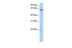
Figure 1. Western blot analysis of LIN9 using anti-LIN9 antibody (A06333-1). Electrophoresis was performed on a 5-20% SDS-PAGE gel at 70V (Stacking gel) / 90V (Resolving gel) for 2-3 hours. The sample well of each lane was loaded with 30 ug of sample under reducing conditions. Lane 1: human 293T whole cell lysates. After electrophoresis, proteins were transferred to a nitrocellulose membrane at 150 mA for 50-90 minutes. Blocked the membrane with 5% non-fat milk/TBS for 1.5 hour at RT. The membrane was incubated with rabbit anti-LIN9 antigen affinity purified polyclonal antibody (Catalog # A06333-1) at 0.5 microg/mL overnight at 4°C, then washed with TBS-0.1%Tween 3 times with 5 minutes each and probed with a goat anti-rabbit IgG-HRP secondary antibody at a dilution of 1:5000 for 1.5 hour at RT. The signal is developed using an Enhanced Chemiluminescent detection (ECL) kit (Catalog # EK1002) with Tanon 5200 system. A specific band was detected for LIN9 at approximately 65 kDa. The expected band size for LIN9 is at 62 kDa.
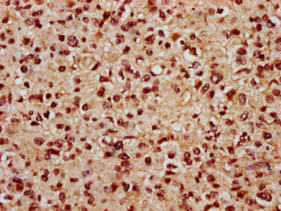
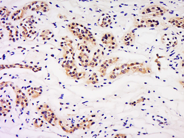
 and anti-Beta Tubulin antibody (M01857-3). LIN9 was detected in immunocytochemical section of HELA cell. Enzyme antigen retrieval was performed using IHC enzyme antigen retrieval reagent (AR0022) for 15 mins. The cells were blocked with 10% goat serum. And then incubated with 5 microg/mL rabbit anti-LIN9 Antibody (A06333-1) and mouse anti-Beta Tubulin antibody (M01857-3) overnight at 4°C. Cy3 Conjugated Goat Anti-Rabbit IgG (BA1032) and DyLight®488 Conjugated Goat Anti-Mouse IgG (BA1126) were used as secondary antibody at 1:500 dilution and incubated for 30 minutes at 37°C. Visualize using a fluorescence microscope and filter sets appropriate for the label used.")
Figure 1. Western blot analysis of LIN9 using anti-LIN9 antibody (A06333-1). Electrophoresis was performed on a 5-20% SDS-PAGE gel at 70V (Stacking gel) / 90V (Resolving gel) for 2-3 hours. The sample well of each lane was loaded with 30 ug of sample under reducing conditions. Lane 1: human 293T whole cell lysates. After electrophoresis, proteins were transferred to a nitrocellulose membrane at 150 mA for 50-90 minutes. Blocked the membrane with 5% non-fat milk/TBS for 1.5 hour at RT. The membrane was incubated with rabbit anti-LIN9 antigen affinity purified polyclonal antibody (Catalog # A06333-1) at 0.5 microg/mL overnight at 4°C, then washed with TBS-0.1%Tween 3 times with 5 minutes each and probed with a goat anti-rabbit IgG-HRP secondary antibody at a dilution of 1:5000 for 1.5 hour at RT. The signal is developed using an Enhanced Chemiluminescent detection (ECL) kit (Catalog # EK1002) with Tanon 5200 system. A specific band was detected for LIN9 at approximately 65 kDa. The expected band size for LIN9 is at 62 kDa.
Anti-LIN9 Antibody Picoband(r)
A06333-1-CARRIER-FREE
ApplicationsImmunoFluorescence, Western Blot, ELISA, ImmunoCytoChemistry
Product group Antibodies
ReactivityHuman
TargetLIN9
Overview
- SupplierBoster Bio
- Product NameAnti-LIN9 Antibody Picoband(r)
- Delivery Days Customer9
- ApplicationsImmunoFluorescence, Western Blot, ELISA, ImmunoCytoChemistry
- CertificationResearch Use Only
- ClonalityPolyclonal
- Concentration500 ug/ml
- Gene ID286826
- Target nameLIN9
- Target descriptionlin-9 DREAM MuvB core complex component
- Target synonymsBARA, BARPsv, Lin-9, TGS, TGS1, TGS2, protein lin-9 homolog, TUDOR gene similar protein, beta subunit-associated regulator of apoptosis, lin-9 homolog, pRB-associated protein, rb related pathway actor, type I interferon receptor beta chain-associated protein
- HostRabbit
- IsotypeIgG
- Protein IDQ5TKA1
- Protein NameProtein lin-9 homolog
- Scientific DescriptionBoster Bio Anti-LIN9 Antibody Picoband® catalog # A06333-1. Tested in ELISA, IF, ICC, WB applications. This antibody reacts with Human. The brand Picoband indicates this is a premium antibody that guarantees superior quality, high affinity, and strong signals with minimal background in Western blot applications. Only our best-performing antibodies are designated as Picoband, ensuring unmatched performance.
- ReactivityHuman
- Storage Instruction-20°C,2°C to 8°C
- UNSPSC12352203
Related products

Product group Antibodies
Anti-LIN9 AntibodyHPA030241
ApplicationsImmunoCytoChemistry, ImmunoHistoChemistry
ReactivityHuman
TargetLIN9
- SizePrice
Product group Antibodies

LIN9 Antibody (FITC)LS-C680772
ApplicationsELISA
ReactivityHuman
TargetLIN9
- SizePrice
Product group Antibodies
LIN9 AntibodyCSB-PA716370LA01HU
ApplicationsELISA, ImmunoHistoChemistry
ReactivityHuman
TargetLIN9
- SizePrice
Product group Antibodies
LIN9 Polyclonal AntibodyBS-18283R
ApplicationsWestern Blot, ELISA
ReactivityHuman, Mouse
TargetLIN9
- SizePrice
Product group Antibodies
LIN9 antibody, N-termGTX49104
ApplicationsWestern Blot
ReactivityHuman
TargetLIN9
- SizePrice
Product group Antibodies
Anti-LIN9 Antibody144-65205
ApplicationsWestern Blot
ReactivityHuman, Mouse
TargetLIN9
- SizePrice