


Immunohistochemical staining of human cerebral cortex shows strong granular cytoplasmic positivity in neurons and neuropil.
Immunohistochemical staining of human cerebral cortex shows strong granular cytoplasmic positivity in neurons and neuropil.
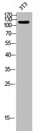
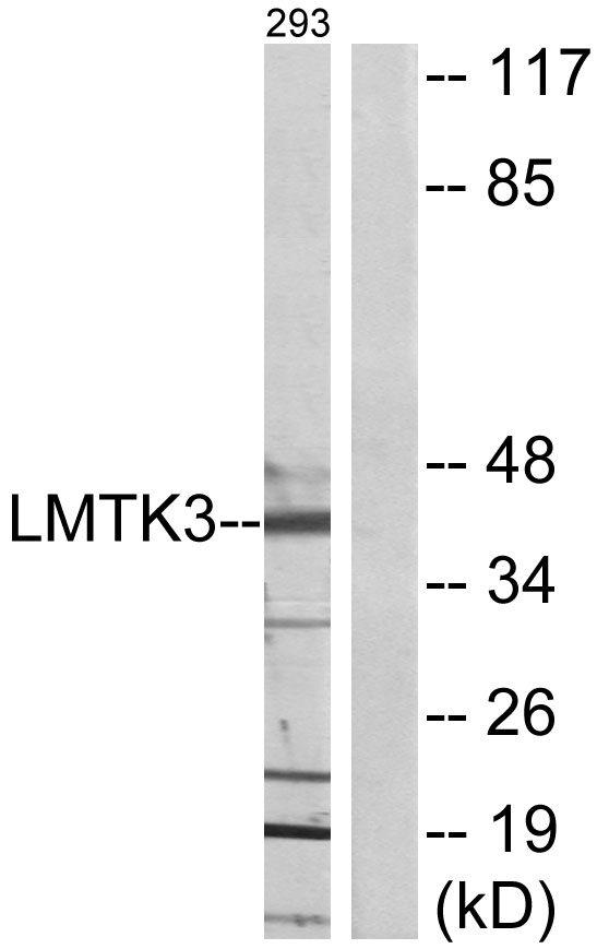
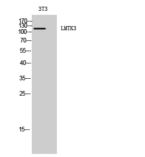
Anti-LMTK3 Antibody
HPA077070
ApplicationsImmunoHistoChemistry
Product group Antibodies
ReactivityHuman, Mouse
TargetLMTK3
Overview
- SupplierAtlas Antibodies
- Product NameAnti-LMTK3 Antibody
- Delivery Days Customer4
- ApplicationsImmunoHistoChemistry
- CertificationResearch Use Only
- ClonalityPolyclonal
- ConjugateUnconjugated
- Gene ID114783
- Target nameLMTK3
- Target descriptionlemur tyrosine kinase 3
- Target synonymsAATYK3, LMR3, PPP1R101, TYKLM3, serine/threonine-protein kinase LMTK3, protein phosphatase 1, regulatory subunit 101
- HostRabbit
- IsotypeIgG
- Protein IDQ96Q04
- Protein NameSerine/threonine-protein kinase LMTK3
- Scientific DescriptionRecombinant Protein Epitope Signature Tag (PrEST) antigen sequence
- ReactivityHuman, Mouse
- Storage Instruction-20°C,2°C to 8°C
- UNSPSC41116161
Datasheet
MSDS
Related products

Product group Antibodies
Anti-LMTK3 Antibody Picoband(r)A08432-2-CARRIER-FREE
ApplicationsWestern Blot, ELISA, ImmunoHistoChemistry
ReactivityHuman, Mouse, Rat
TargetLMTK3
- SizePrice
Product group Antibodies
Anti-LMTK3 Antibody101-10923
ApplicationsWestern Blot, ELISA
TargetLMTK3
- SizePrice
Product group Antibodies
LMTK3 AntibodyCSB-PA006834
ApplicationsWestern Blot, ELISA, ImmunoHistoChemistry
ReactivityHuman, Mouse
- SizePrice
Product group Antibodies
Anti-LMTK3 AntibodyA99052
ApplicationsWestern Blot, ELISA
ReactivityHuman, Mouse
- SizePrice
Product group Antibodies
LMTK3 antibodyGTX34041
ApplicationsWestern Blot
ReactivityHuman, Mouse
TargetLMTK3
- SizePrice
Product group Antibodies

LMTK3 Antibody (C-Terminus)LS-C358796
ApplicationsWestern Blot, ImmunoHistoChemistry, ImmunoHistoChemistry Paraffin
ReactivityHuman, Mouse
TargetLMTK3
- SizePrice