

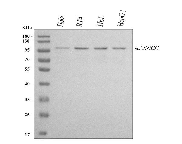
Figure 1. Western blot analysis of LONRF1 using anti-LONRF1 antibody (A14796). Electrophoresis was performed on a 5-20% SDS-PAGE gel at 70V (Stacking gel) / 90V (Resolving gel) for 2-3 hours. The sample well of each lane was loaded with 30 ug of sample under reducing conditions. Lane 1: human Hela whole cell lysates, Lane 2: human RT4 whole cell lysates, Lane 3: human HEL whole cell lysates, Lane 4: human HepG2 whole cell lysates. After electrophoresis, proteins were transferred to a nitrocellulose membrane at 150 mA for 50-90 minutes. Blocked the membrane with 5% non-fat milk/TBS for 1.5 hour at RT. The membrane was incubated with rabbit anti-LONRF1 antigen affinity purified polyclonal antibody (Catalog # A14796) at 0.5 microg/mL overnight at 4°C, then washed with TBS-0.1%Tween 3 times with 5 minutes each and probed with a goat anti-rabbit IgG-HRP secondary antibody at a dilution of 1:5000 for 1.5 hour at RT. The signal is developed using an Enhanced Chemiluminescent detection (ECL) kit (Catalog # EK1002) with Tanon 5200 system. A specific band was detected for LONRF1 at approximately 100 kDa. The expected band size for LONRF1 is at 87 kDa.
Figure 1. Western blot analysis of LONRF1 using anti-LONRF1 antibody (A14796). Electrophoresis was performed on a 5-20% SDS-PAGE gel at 70V (Stacking gel) / 90V (Resolving gel) for 2-3 hours. The sample well of each lane was loaded with 30 ug of sample under reducing conditions. Lane 1: human Hela whole cell lysates, Lane 2: human RT4 whole cell lysates, Lane 3: human HEL whole cell lysates, Lane 4: human HepG2 whole cell lysates. After electrophoresis, proteins were transferred to a nitrocellulose membrane at 150 mA for 50-90 minutes. Blocked the membrane with 5% non-fat milk/TBS for 1.5 hour at RT. The membrane was incubated with rabbit anti-LONRF1 antigen affinity purified polyclonal antibody (Catalog # A14796) at 0.5 microg/mL overnight at 4°C, then washed with TBS-0.1%Tween 3 times with 5 minutes each and probed with a goat anti-rabbit IgG-HRP secondary antibody at a dilution of 1:5000 for 1.5 hour at RT. The signal is developed using an Enhanced Chemiluminescent detection (ECL) kit (Catalog # EK1002) with Tanon 5200 system. A specific band was detected for LONRF1 at approximately 100 kDa. The expected band size for LONRF1 is at 87 kDa.
Anti-LONRF1 Antibody Picoband(r)
A14796-CARRIER-FREE
ApplicationsWestern Blot, ELISA
Product group Antibodies
ReactivityHuman
TargetLONRF1
Overview
- SupplierBoster Bio
- Product NameAnti-LONRF1 Antibody Picoband(r)
- Delivery Days Customer9
- Application Supplier NoteTested Species: In-house tested species with positive results. Other applications have not been tested. Optimal dilutions should be determined by end users.
- ApplicationsWestern Blot, ELISA
- CertificationResearch Use Only
- ClonalityPolyclonal
- Concentration500 ug/ml
- Gene ID91694
- Target nameLONRF1
- Target descriptionLON peptidase N-terminal domain and ring finger 1
- Target synonymsRNF191, LON peptidase N-terminal domain and RING finger protein 1, RING finger protein 191
- HostRabbit
- IsotypeIgG
- Protein IDQ17RB8
- Protein NameLON peptidase N-terminal domain and RING finger protein 1
- Scientific DescriptionBoster Bio Anti-LONRF1 Antibody Picoband® catalog # A14796. Tested in ELISA, WB applications. This antibody reacts with Human. The brand Picoband indicates this is a premium antibody that guarantees superior quality, high affinity, and strong signals with minimal background in Western blot applications. Only our best-performing antibodies are designated as Picoband, ensuring unmatched performance.
- ReactivityHuman
- Storage Instruction-20°C,2°C to 8°C
- UNSPSC12352203
Related products

Product group Antibodies
Anti-LONRF1 Antibody101-10928
ApplicationsWestern Blot, ELISA
TargetLONRF1
- SizePrice
Product group Antibodies
LONRF1 AntibodyCSB-PA615067LA01HU
ApplicationsELISA, ImmunoHistoChemistry
ReactivityHuman
TargetLONRF1
- SizePrice
Product group Antibodies

LONRF1 AntibodyLS-C749434
ApplicationsWestern Blot
ReactivityHuman, Mouse, Rat
TargetLONRF1
- SizePrice
Product group Antibodies
LONRF1 antibody, N-termGTX46891
ApplicationsWestern Blot, ImmunoHistoChemistry, ImmunoHistoChemistry Paraffin
ReactivityHuman
TargetLONRF1
- SizePrice

Product group Antibodies
Anti-LONRF1 AntibodyHPA023584
ApplicationsImmunoCytoChemistry, ImmunoHistoChemistry
ReactivityHuman
TargetLONRF1
- SizePrice