

Anti-Low Density Lipoprotein Receptor (LDLR) Polyclonal Antibody
CAU35506
ApplicationsFlow Cytometry
Product group Antibodies
TargetLDLR
Overview
- SupplierBiomatik
- Product NameAnti-Low Density Lipoprotein Receptor (LDLR) Polyclonal Antibody
- Delivery Days Customer12
- ApplicationsFlow Cytometry
- CertificationResearch Use Only
- ClonalityPolyclonal
- Concentration0.5 mg/ml
- ConjugateUnconjugated
- Gene ID3949
- Target nameLDLR
- Target descriptionlow density lipoprotein receptor
- Target synonymsFH, FHC, FHCL1, LDLCQ2, low-density lipoprotein receptor, LDL receptor, low-density lipoprotein receptor class A domain-containing protein 3
- HostRabbit
- Protein IDP01130
- Protein NameLow-density lipoprotein receptor
- Scientific DescriptionThe Anti-Low Density Lipoprotein Receptor (LDLR) Polyclonal Antibody (Species: Human) has been validated for the following applications: Flow cytometry.
- Storage Instruction-20°C,2°C to 8°C
- UNSPSC12352203
Related products
Product group Antibodies
Anti-LDL Receptor AntibodyA308995
ApplicationsImmunoFluorescence, Western Blot, ImmunoCytoChemistry, ImmunoHistoChemistry
ReactivityHuman, Mouse, Rat
- SizePrice
Product group Antibodies

Anti-LDL Receptor [C7]AB03080-2.0
ApplicationsFlow Cytometry, ImmunoFluorescence, ImmunoPrecipitation, Western Blot, Neutralisation/Blocking
ReactivityBovine, Human
TargetLDLR
- SizePrice
Product group Antibodies
Anti-LDLR Antibody144-60878
ApplicationsWestern Blot
ReactivityHuman, Mouse
TargetLDLR
- SizePrice

Product group Antibodies
Anti-LDL Receptor/LDLR Antibody Picoband(r)A00076-2-CARRIER-FREE
ApplicationsFlow Cytometry, Western Blot, ELISA, ImmunoHistoChemistry
ReactivityHuman, Rat
TargetLDLR
- SizePrice
Product group Antibodies
References
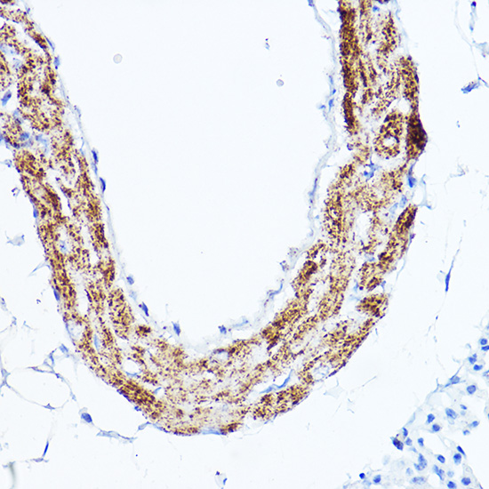
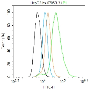
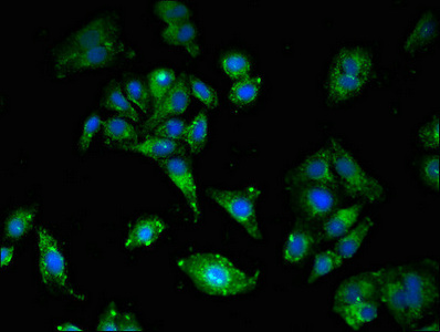
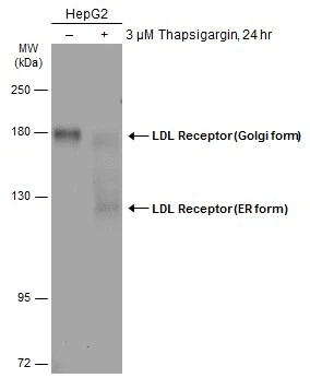
LDLR Polyclonal AntibodyBS-0705R
ApplicationsFlow Cytometry, ImmunoFluorescence, Western Blot, ELISA, ImmunoHistoChemistry, ImmunoHistoChemistry Paraffin
ReactivityBovine, Canine, Equine, Guinea Pig, Hamster, Human, Mouse, Porcine, Rabbit, Rat
TargetLDLR
- SizePrice
Product group Antibodies
LDLR AntibodyCSB-PA09949A0RB
ApplicationsImmunoFluorescence, ELISA
ReactivityHuman
TargetLDLR
- SizePrice
Product group Antibodies
LDL Receptor antibodyGTX132860
ApplicationsWestern Blot
ReactivityHuman, Rat
TargetLDLR
- SizePrice
Product group Antibodies

LDLR / LDL Receptor Antibody (APC)LS-C488074
ApplicationsFlow Cytometry
ReactivityHuman
TargetLDLR
- SizePrice

Product group Antibodies
Anti-LDLR AntibodyHPA009647
ApplicationsImmunoHistoChemistry
ReactivityHuman
TargetLDLR
- SizePrice