


Figure 1. Western blot analysis of LRRC75B using anti-LRRC75B antibody (A18683-1). Electrophoresis was performed on a 5-20% SDS-PAGE gel at 70V (Stacking gel) / 90V (Resolving gel) for 2-3 hours. The sample well of each lane was loaded with 30 ug of sample under reducing conditions. Lane 1: human HepG2 whole cell lysates, Lane 2: human SH-SY5Y whole cell lysates, Lane 3: human MCF-7 whole cell lysates, Lane 4: rat C6 whole cell lysates, Lane 5: mouse brain tissue lysates, Lane 6: mouse Neuro-2a whole cell lysates. After electrophoresis, proteins were transferred to a nitrocellulose membrane at 150 mA for 50-90 minutes. Blocked the membrane with 5% non-fat milk/TBS for 1.5 hour at RT. The membrane was incubated with rabbit anti-LRRC75B antigen affinity purified polyclonal antibody (Catalog # A18683-1) at 0.5 microg/mL overnight at 4°C, then washed with TBS-0.1%Tween 3 times with 5 minutes each and probed with a goat anti-rabbit IgG-HRP secondary antibody at a dilution of 1:5000 for 1.5 hour at RT. The signal is developed using an Enhanced Chemiluminescent detection (ECL) kit (Catalog # EK1002) with Tanon 5200 system. A specific band was detected for LRRC75B at approximately 35 kDa. The expected band size for LRRC75B is at 35 kDa.
. LRRC75B was detected in a paraffin-embedded section of human colorectal adenocarcinoma tissue. Heat mediated antigen retrieval was performed in EDTA buffer (pH 8.0, epitope retrieval solution). The tissue section was blocked with 10% goat serum. The tissue section was then incubated with 2 microg/ml rabbit anti-LRRC75B Antibody (A18683-1) overnight at 4°C. Peroxidase Conjugated Goat Anti-rabbit IgG was used as secondary antibody and incubated for 30 minutes at 37°C. The tissue section was developed using HRP Conjugated Rabbit IgG Super Vision Assay Kit (Catalog # SV0002) with DAB as the chromogen.")
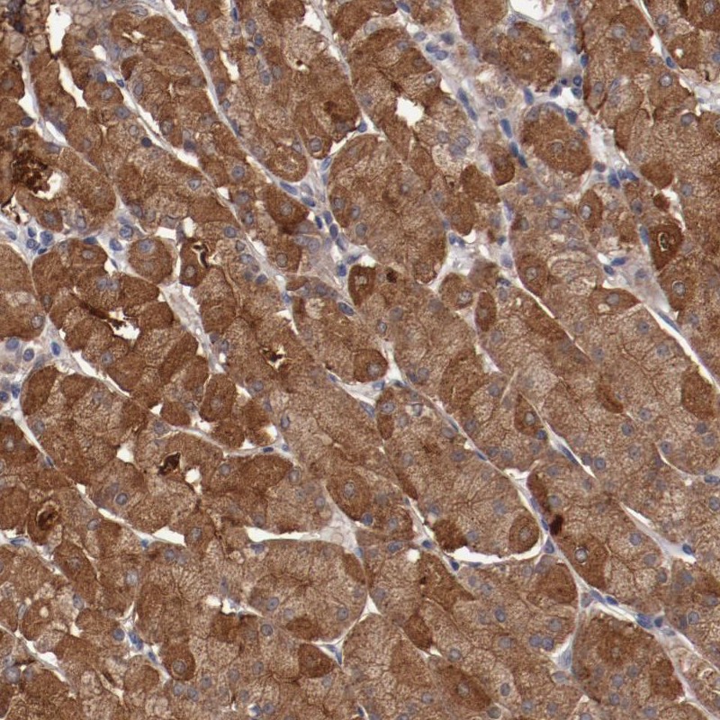
. LRRC75B was detected in a paraffin-embedded section of human liver cancer tissue. Heat mediated antigen retrieval was performed in EDTA buffer (pH 8.0, epitope retrieval solution). The tissue section was blocked with 10% goat serum. The tissue section was then incubated with 2 microg/ml rabbit anti-LRRC75B Antibody (A18683-1) overnight at 4°C. Peroxidase Conjugated Goat Anti-rabbit IgG was used as secondary antibody and incubated for 30 minutes at 37°C. The tissue section was developed using HRP Conjugated Rabbit IgG Super Vision Assay Kit (Catalog # SV0002) with DAB as the chromogen.")
. LRRC75B was detected in a paraffin-embedded section of human lung cancer tissue. Heat mediated antigen retrieval was performed in EDTA buffer (pH 8.0, epitope retrieval solution). The tissue section was blocked with 10% goat serum. The tissue section was then incubated with 2 microg/ml rabbit anti-LRRC75B Antibody (A18683-1) overnight at 4°C. Peroxidase Conjugated Goat Anti-rabbit IgG was used as secondary antibody and incubated for 30 minutes at 37°C. The tissue section was developed using HRP Conjugated Rabbit IgG Super Vision Assay Kit (Catalog # SV0002) with DAB as the chromogen.")
. Overlay histogram showing PC-3 cells stained with A18683-1 (Blue line). To facilitate intracellular staining, cells were fixed with 4% paraformaldehyde and permeabilized with permeabilization buffer. The cells were blocked with 10% normal goat serum. And then incubated with rabbit anti-LRRC75B Antibody (A18683-1, 1 microg/1x106 cells) for 30 min at 20°C. DyLight®488 conjugated goat anti-rabbit IgG (BA1127, 5-10 microg/1x106 cells) was used as secondary antibody for 30 minutes at 20°C. Isotype control antibody (Green line) was rabbit IgG (1 microg/1x106) used under the same conditions. Unlabelled sample (Red line) was also used as a control.")
Figure 1. Western blot analysis of LRRC75B using anti-LRRC75B antibody (A18683-1). Electrophoresis was performed on a 5-20% SDS-PAGE gel at 70V (Stacking gel) / 90V (Resolving gel) for 2-3 hours. The sample well of each lane was loaded with 30 ug of sample under reducing conditions. Lane 1: human HepG2 whole cell lysates, Lane 2: human SH-SY5Y whole cell lysates, Lane 3: human MCF-7 whole cell lysates, Lane 4: rat C6 whole cell lysates, Lane 5: mouse brain tissue lysates, Lane 6: mouse Neuro-2a whole cell lysates. After electrophoresis, proteins were transferred to a nitrocellulose membrane at 150 mA for 50-90 minutes. Blocked the membrane with 5% non-fat milk/TBS for 1.5 hour at RT. The membrane was incubated with rabbit anti-LRRC75B antigen affinity purified polyclonal antibody (Catalog # A18683-1) at 0.5 microg/mL overnight at 4°C, then washed with TBS-0.1%Tween 3 times with 5 minutes each and probed with a goat anti-rabbit IgG-HRP secondary antibody at a dilution of 1:5000 for 1.5 hour at RT. The signal is developed using an Enhanced Chemiluminescent detection (ECL) kit (Catalog # EK1002) with Tanon 5200 system. A specific band was detected for LRRC75B at approximately 35 kDa. The expected band size for LRRC75B is at 35 kDa.
Anti-LRRC75B Antibody Picoband(r)
A18683-1-BIOTIN
ApplicationsFlow Cytometry, Western Blot, ELISA, ImmunoHistoChemistry
Product group Antibodies
ReactivityHuman, Mouse, Rat
TargetLRRC75B
Overview
- SupplierBoster Bio
- Product NameAnti-LRRC75B Antibody Picoband(r)
- Delivery Days Customer9
- ApplicationsFlow Cytometry, Western Blot, ELISA, ImmunoHistoChemistry
- CertificationResearch Use Only
- ClonalityPolyclonal
- Concentration500 ug/ml
- ConjugateBiotin
- Gene ID388886
- Target nameLRRC75B
- Target descriptionleucine rich repeat containing 75B
- Target synonymsC22orf36, FAM211B, leucine-rich repeat-containing protein 75B, family with sequence similarity 211, member B, leucine-rich repeat-containing protein C22orf36, leucine-rich repeat-containing protein FAM211B
- HostRabbit
- IsotypeIgG
- Protein IDQ2VPJ9
- Protein NameLeucine-rich repeat-containing protein 75B
- Scientific DescriptionBoster Bio Anti-LRRC75B Antibody Picoband® catalog # A18683-1. Tested in ELISA, IHC, WB, Flow Cytometry applications. This antibody reacts with Human, Mouse, Rat. The brand Picoband indicates this is a premium antibody that guarantees superior quality, high affinity, and strong signals with minimal background in Western blot applications. Only our best-performing antibodies are designated as Picoband, ensuring unmatched performance.
- ReactivityHuman, Mouse, Rat
- Storage Instruction-20°C,2°C to 8°C
- UNSPSC12352203
Related products
Product group Antibodies

LRRC75B / FAM211B Antibody (aa107-156)LS-C117187
ApplicationsWestern Blot
ReactivityHuman
TargetLRRC75B
- SizePrice
Product group Antibodies
Anti-LRRC75B AntibodyHPA002540
ApplicationsImmunoHistoChemistry
ReactivityHuman
TargetLRRC75B
- SizePrice
Product group Antibodies
Anti-LRRC75B Antibody Picoband(r)A18683-1-CARRIER-FREE
ApplicationsFlow Cytometry, Western Blot, ELISA, ImmunoHistoChemistry
ReactivityHuman, Mouse, Rat
TargetLRRC75B
- SizePrice