

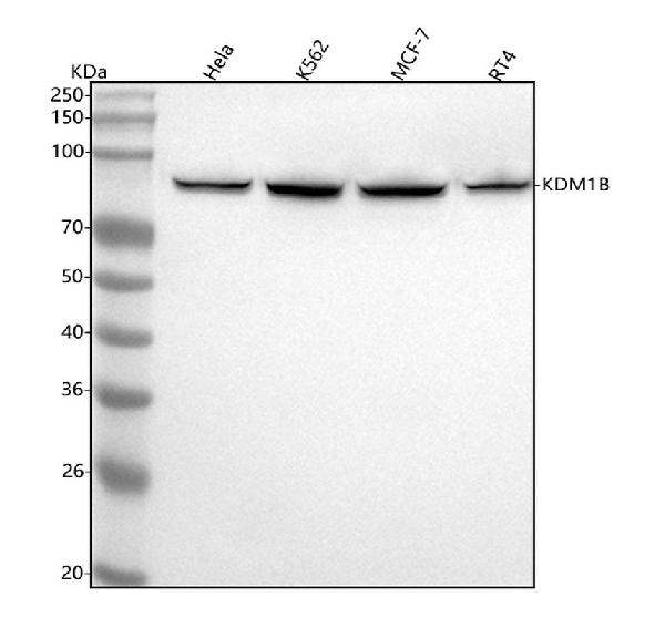
Figure 1. Western blot analysis of KDM1B using anti-KDM1B antibody (M08071). Electrophoresis was performed on a 5-20% SDS-PAGE gel at 70V (Stacking gel) / 90V (Resolving gel) for 2-3 hours. The sample well of each lane was loaded with 30 ug of sample under reducing conditions. Lane 1: human Hela whole cell lysates, Lane 2: human K562 whole cell lysates, Lane 3: human MCF-7 whole cell lysates, Lane 4: human RT4 whole cell lysates. After electrophoresis, proteins were transferred to a nitrocellulose membrane at 150 mA for 50-90 minutes. Blocked the membrane with 5% non-fat milk/TBS for 1.5 hour at RT. The membrane was incubated with rabbit anti-KDM1B antigen affinity purified monoclonal antibody (Catalog # M08071) at 1:500 overnight at 4°C, then washed with TBS-0.1%Tween 3 times with 5 minutes each and probed with a goat anti-rabbit IgG-HRP secondary antibody at a dilution of 1:500 for 1.5 hour at RT. The signal is developed using an Enhanced Chemiluminescent detection (ECL) kit (Catalog # EK1002) with Tanon 5200 system. A specific band was detected for KDM1B at approximately 93 kDa. The expected band size for KDM1B is at 93 kDa.
 HeLa cell lysate; (2) RAW264.7 cell lysate; (3) PC12 cell lysate.")
Figure 1. Western blot analysis of KDM1B using anti-KDM1B antibody (M08071). Electrophoresis was performed on a 5-20% SDS-PAGE gel at 70V (Stacking gel) / 90V (Resolving gel) for 2-3 hours. The sample well of each lane was loaded with 30 ug of sample under reducing conditions. Lane 1: human Hela whole cell lysates, Lane 2: human K562 whole cell lysates, Lane 3: human MCF-7 whole cell lysates, Lane 4: human RT4 whole cell lysates. After electrophoresis, proteins were transferred to a nitrocellulose membrane at 150 mA for 50-90 minutes. Blocked the membrane with 5% non-fat milk/TBS for 1.5 hour at RT. The membrane was incubated with rabbit anti-KDM1B antigen affinity purified monoclonal antibody (Catalog # M08071) at 1:500 overnight at 4°C, then washed with TBS-0.1%Tween 3 times with 5 minutes each and probed with a goat anti-rabbit IgG-HRP secondary antibody at a dilution of 1:500 for 1.5 hour at RT. The signal is developed using an Enhanced Chemiluminescent detection (ECL) kit (Catalog # EK1002) with Tanon 5200 system. A specific band was detected for KDM1B at approximately 93 kDa. The expected band size for KDM1B is at 93 kDa.
Anti-LSD2 / AOF1 Monoclonal Antibody
M08071
ApplicationsFlow Cytometry, ImmunoFluorescence, ImmunoPrecipitation, Western Blot, ImmunoCytoChemistry
Product group Antibodies
ReactivityHuman, Mouse, Rat
TargetKDM1B
Overview
- SupplierBoster Bio
- Product NameAnti-LSD2 / AOF1 Monoclonal Antibody
- Delivery Days Customer9
- ApplicationsFlow Cytometry, ImmunoFluorescence, ImmunoPrecipitation, Western Blot, ImmunoCytoChemistry
- CertificationResearch Use Only
- ClonalityMonoclonal
- Clone IDADAG-11
- Gene ID221656
- Target nameKDM1B
- Target descriptionlysine demethylase 1B
- Target synonymsAOF1, C6orf193, LSD2, lysine-specific histone demethylase 2, flavin-containing amine oxidase domain-containing protein 1, lysine (K)-specific demethylase 1B, lysine-specific histone demethylase 1B
- HostRabbit
- IsotypeIgG
- Protein IDQ8NB78
- Protein NameLysine-specific histone demethylase 2
- Scientific DescriptionBoster Bio Anti-LSD2 / AOF1 Monoclonal Antibody catalog # M08071. Tested in WB, ICC/IF, IP, Flow Cytometry applications. This antibody reacts with Human, Mouse, Rat.
- ReactivityHuman, Mouse, Rat
- Storage Instruction-20°C
- UNSPSC12352203
Datasheet
MSDS
Related products
Product group Antibodies

KDM1B AntibodyLS-C678413
ApplicationsELISA, ImmunoHistoChemistry, ImmunoHistoChemistry Paraffin
ReactivityHuman
TargetKDM1B
- SizePrice
Product group Antibodies
KDM1B AntibodyCSB-PA822748LA01HU
ApplicationsELISA, ImmunoHistoChemistry
ReactivityHuman
TargetKDM1B
- SizePrice
Product group Antibodies
KDM1B Recombinant Antibody, AbBy Fluor-594 ConjugatedBSM-61638R-BF594
ApplicationsFlow Cytometry, Western Blot
ReactivityHuman, Mouse, Rat
TargetKDM1B
- SizePrice
Product group Antibodies
Kdm1B Recombinant AntibodyCAC12305
ApplicationsWestern Blot, ELISA
TargetKDM1B
- SizePrice
![Various whole cell extracts (30 μg) were separated by 7.5% SDS-PAGE, and the membrane was blotted with LSD2 / AOF1 antibody [HL3728] (GTX641918) diluted at 1:1000. The HRP-conjugated anti-rabbit IgG antibody (GTX213110-01) was used to detect the primary antibody.](https://www.genetex.com/upload/website/prouct_img/normal/GTX641918/GTX641918_T-45663_20250124_WB_25020422_420.webp)
Product group Antibodies
LSD2 / AOF1 antibody [HL3728]GTX641918
ApplicationsWestern Blot
ReactivityHuman
TargetKDM1B
- SizePrice
Product group Antibodies
Anti-KDM1B (N-term) Antibody102-24201
ApplicationsWestern Blot
TargetKDM1B
- SizePrice