


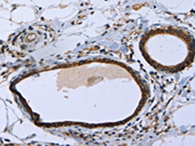
Immunohistochemical staining of human cerebral cortex shows strong cytoplasmic positivity in neuronal cells.

Immunohistochemical staining of human cerebral cortex shows strong cytoplasmic positivity in neuronal cells.
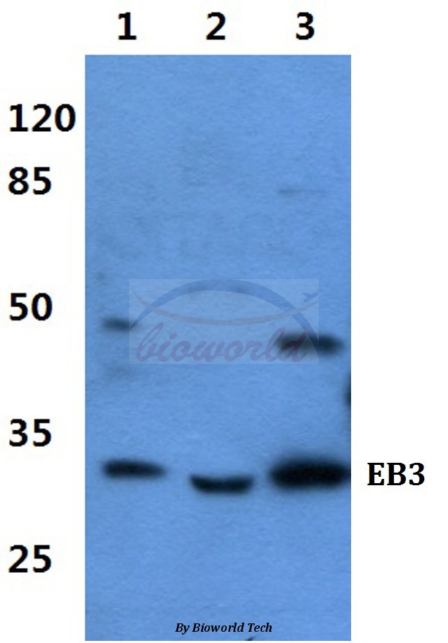
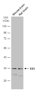
Anti-MAPRE3 Antibody
HPA009263
ApplicationsWestern Blot, ImmunoHistoChemistry
Product group Antibodies
ReactivityHuman
TargetMAPRE3
Overview
- SupplierAtlas Antibodies
- Product NameAnti-MAPRE3 Antibody
- Delivery Days Customer4
- ApplicationsWestern Blot, ImmunoHistoChemistry
- CertificationResearch Use Only
- ClonalityPolyclonal
- ConjugateUnconjugated
- Gene ID22924
- Target nameMAPRE3
- Target descriptionmicrotubule associated protein RP/EB family member 3
- Target synonymsEB3, EBF3, EBF3-S, RP3, microtubule-associated protein RP/EB family member 3, APC binding protein, EB1 protein family member 3, end-binding protein 3
- HostRabbit
- IsotypeIgG
- Protein IDQ9UPY8
- Protein NameMicrotubule-associated protein RP/EB family member 3
- Scientific DescriptionRecombinant Protein Epitope Signature Tag (PrEST) antigen sequence
- ReactivityHuman
- Storage Instruction-20°C,2°C to 8°C
- UNSPSC41116161
Datasheet
MSDS
Related products
Product group Antibodies
Anti-EB3 AntibodyA28608
ApplicationsWestern Blot
ReactivityHuman, Mouse, Rat
- SizePrice

Product group Antibodies
Anti-EB3/MAPRE3 Antibody Picoband(r)A05782-2-CARRIER-FREE
ApplicationsFlow Cytometry, Western Blot, ELISA
ReactivityHuman, Mouse, Rat
TargetMAPRE3
- SizePrice
Product group Antibodies
Anti-MAPRE3 Antibody144-09591
ApplicationsWestern Blot, ImmunoHistoChemistry
ReactivityHuman, Mouse
TargetMAPRE3
- SizePrice
Product group Antibodies
MAPRE3 Recombinant Antibody, AbBy Fluor-405 ConjugatedBSM-61869R-BF405
ApplicationsFlow Cytometry, ImmunoFluorescence, Western Blot
ReactivityHuman, Mouse, Rat
TargetMAPRE3
- SizePrice
Product group Antibodies

Goat anti-MAPRE3EB09827
ApplicationsWestern Blot, ELISA
ReactivityBovine, Canine, Human, Mouse
TargetMAPRE3
- SizePrice
Product group Antibodies
MAPRE3 AntibodyCSB-PA059696
ApplicationsWestern Blot, ELISA, ImmunoHistoChemistry
ReactivityHuman, Mouse, Rat
TargetMAPRE3
- SizePrice
Product group Antibodies

MAPRE3 / EB3 AntibodyLS-C401618
ApplicationsWestern Blot, ELISA, ImmunoHistoChemistry
ReactivityHuman, Mouse, Rat
TargetMAPRE3
- SizePrice
Product group Antibodies
EB3 antibodyGTX115652
ApplicationsWestern Blot, ImmunoHistoChemistry, ImmunoHistoChemistry Paraffin
ReactivityHuman, Mouse, Rat
TargetMAPRE3
- SizePrice