

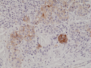
Immunohistochemical staining of formalin fixed and paraffin embedded human melanoma tissue section using anti-MART1 rabbit monoclonal antibody (Clone RM333) at a 1:1000 dilution.
Immunohistochemical staining of formalin fixed and paraffin embedded human melanoma tissue section using anti-MART1 rabbit monoclonal antibody (Clone RM333) at a 1:1000 dilution.
anti-MART1 (human), Rabbit Monoclonal (RM333)
REV-31-1220-00
ApplicationsWestern Blot, ImmunoHistoChemistry
Product group Antibodies
ReactivityHuman
TargetMLANA
Overview
- SupplierRevMAb Biosciences
- Product Nameanti-MART1 (human), Rabbit Monoclonal (RM333)
- Delivery Days Customer2
- ApplicationsWestern Blot, ImmunoHistoChemistry
- CertificationResearch Use Only
- ClonalityMonoclonal
- Clone IDRM333
- Gene ID2315
- Target nameMLANA
- Target descriptionmelan-A
- Target synonymsMART-1, MART1, melanoma antigen recognized by T-cells 1, antigen LB39-AA, antigen SK29-AA, epididymis secretory sperm binding protein, protein Melan-A
- HostRabbit
- IsotypeIgG
- Protein IDQ16655
- Protein NameMelanoma antigen recognized by T-cells 1
- Scientific DescriptionMART1 is involved in melanosome biogenesis by ensuring the stability of GPR143. It plays a vital role in the expression, stability, trafficking and processing of melanocyte protein PMEL, which is critical to the formation of stage II melanosomes. MART1 is found on the surface of melanocytes and is specific for the melanocyte lineage, found in normal skin, the retina and melanocytes, but not in other normal tissues. It is thus useful as a marker for melanocytic tumors (melanomas). - Recombinant Antibody. This antibody reacts to human MART1 (Melanoma antigen recognized by T-cells-1). Applications: WB, IHC. Source: Rabbit. Liquid. 50% Glycerol/PBS with 1% BSA and 0.09% sodium azide. MART1 is involved in melanosome biogenesis by ensuring the stability of GPR143. It plays a vital role in the expression, stability, trafficking and processing of melanocyte protein PMEL, which is critical to the formation of stage II melanosomes. MART1 is found on the surface of melanocytes and is specific for the melanocyte lineage, found in normal skin, the retina and melanocytes, but not in other normal tissues. It is thus useful as a marker for melanocytic tumors (melanomas).
- ReactivityHuman
- Storage Instruction-20°C,2°C to 8°C
- UNSPSC41116161
Datasheet
Related products
Product group Antibodies
MLANA AntibodyCSB-PA008328
ApplicationsWestern Blot, ELISA, ImmunoHistoChemistry
ReactivityHuman
TargetMLANA
- SizePrice

Product group Antibodies
Anti-Melan-A/MLANA Antibody Picoband(r)A02033-3-CARRIER-FREE
ApplicationsFlow Cytometry, Western Blot, ELISA
ReactivityHuman, Mouse
TargetMLANA
- SizePrice
Product group Antibodies

MART-1 AntibodyABX013135
ApplicationsWestern Blot, ELISA, ImmunoHistoChemistry
- SizePrice

Product group Antibodies
Anti-MLANA AntibodyAMAB91816
ApplicationsWestern Blot, ImmunoCytoChemistry, ImmunoHistoChemistry
ReactivityHuman
TargetMLANA
- SizePrice
Product group Antibodies

Anti-Melan-A [A103]Ab02840-1.1
ApplicationsImmunoPrecipitation, Western Blot, ELISA, ImmunoHistoChemistry
ReactivityHuman
TargetMLANA
- SizePrice
Product group Antibodies
Anti-MART-1 AntibodyA99292
ApplicationsWestern Blot, ELISA, ImmunoHistoChemistry
ReactivityHuman
- SizePrice
Product group Antibodies

MLANA / Melan-A AntibodyLS-C831121
ApplicationsELISA, ImmunoHistoChemistry
ReactivityHuman
TargetMLANA
- SizePrice
Product group Antibodies
MLANA Polyclonal AntibodyCAC14190
ApplicationsWestern Blot, ELISA, ImmunoHistoChemistry
TargetMLANA
- SizePrice

Product group Antibodies
Melan A Recombinant AntibodyBSM-52337R
ApplicationsFlow Cytometry, ImmunoFluorescence, Western Blot, ImmunoCytoChemistry, ImmunoHistoChemistry, ImmunoHistoChemistry Frozen, ImmunoHistoChemistry Paraffin
ReactivityHuman
TargetMLANA
- SizePrice