


Figure 2. Immunofluorescent staining data of MATN1 using Anti-Matrilin-1 MATN1 Monoclonal Antibody (M07385). Immunofluorescence (IF) analysis of HeLa cells using Matrilin-1 Monoclonal Antibody (green). Blue: DRAQ5 fluorescent DNA dye. Red: Actin filaments have been labeled with Alexa Fluor-555 phalloidin.
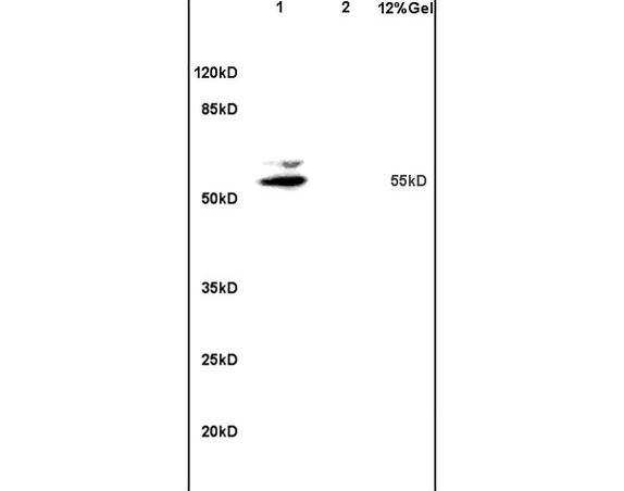
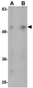
 analysis using Matrilin-1 Monoclonal Antibody against HEK293 (1) and MATN1-hIgGFc transfected HEK293 (2) cell lysate. Electrophoresis was performed on a SDS-PAGE gel. To determine SDS-PAGE gel concentration")
Figure 2. Immunofluorescent staining data of MATN1 using Anti-Matrilin-1 MATN1 Monoclonal Antibody (M07385). Immunofluorescence (IF) analysis of HeLa cells using Matrilin-1 Monoclonal Antibody (green). Blue: DRAQ5 fluorescent DNA dye. Red: Actin filaments have been labeled with Alexa Fluor-555 phalloidin.
Anti-Matrilin-1 MATN1 Monoclonal Antibody
M07385
ApplicationsImmunoFluorescence, Western Blot, ELISA
Product group Antibodies
ReactivityHuman
TargetMATN1
Overview
- SupplierBoster Bio
- Product NameAnti-Matrilin-1 MATN1 Monoclonal Antibody
- Delivery Days Customer9
- ApplicationsImmunoFluorescence, Western Blot, ELISA
- CertificationResearch Use Only
- ClonalityMonoclonal
- Clone ID5A8
- Concentration1 mg/ml
- Gene ID4146
- Target nameMATN1
- Target descriptionmatrilin 1
- Target synonymsCMP, CRTM, matrilin-1, matrilin 1, cartilage matrix protein
- HostMouse
- IsotypeIgG
- Protein IDP21941
- Protein NameMatrilin-1
- Scientific DescriptionBoster Bio Anti-Matrilin-1 MATN1 Monoclonal Antibody catalog # M07385. Tested in WB, IF, ELISA applications. This antibody reacts with Human.
- ReactivityHuman
- Storage Instruction-20°C,2°C to 8°C
- UNSPSC12352203
Related products
Product group Antibodies
MATN1 AntibodyCSB-PA602770
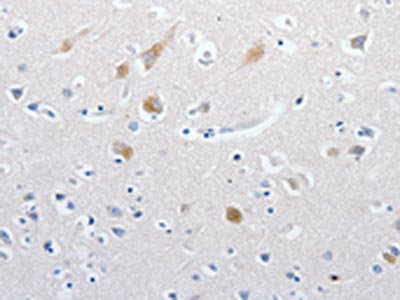
ApplicationsELISA, ImmunoHistoChemistry
ReactivityHuman
TargetMATN1
- SizePrice
Product group Antibodies
Anti-MATN1 AntibodyA46102
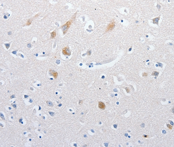
ApplicationsImmunoHistoChemistry
ReactivityHuman
- SizePrice
Product group Antibodies
Anti-MATN1 AntibodyHPA028580
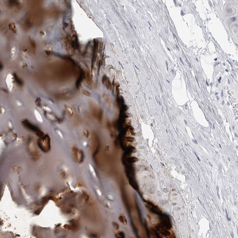
ApplicationsImmunoHistoChemistry
ReactivityHuman
TargetMATN1
- SizePrice
Product group Antibodies

MATN1 / Matrilin 1 AntibodyLS-C405706
ApplicationsELISA, ImmunoHistoChemistry
ReactivityHuman
TargetMATN1
- SizePrice
Product group Antibodies
MATN1 AntibodyPACO18189
ApplicationsELISA, ImmunoHistoChemistry
ReactivityHuman
TargetMATN1
- SizePrice
Product group Antibodies
Matrilin 1 Polyclonal AntibodyBS-1976R
ApplicationsImmunoFluorescence, Western Blot, ELISA, ImmunoCytoChemistry, ImmunoHistoChemistry, ImmunoHistoChemistry Frozen, ImmunoHistoChemistry Paraffin
ReactivityCanine, Equine, Human, Mouse, Rabbit, Rat
TargetMATN1
- SizePrice
Product group Antibodies
MATN1 antibodyGTX85287
ApplicationsWestern Blot, ELISA
ReactivityHuman, Mouse, Rat
TargetMATN1
- SizePrice