

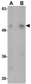
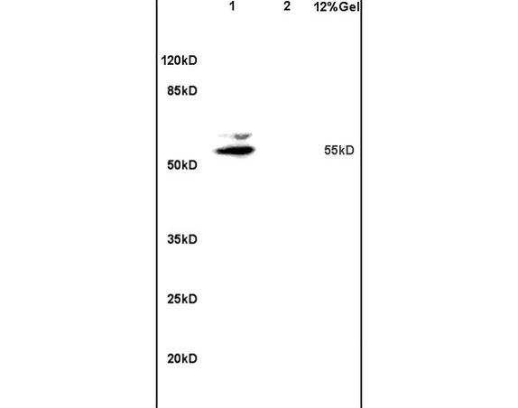
WB analysis of mouse liver tissue lysate using GTX85287 MATN1 antibody. Working concentration : (A) 1 and (B) 2 μg/ml
WB analysis of mouse liver tissue lysate using GTX85287 MATN1 antibody. Working concentration : (A) 1 and (B) 2 μg/ml
MATN1 antibody
GTX85287
ApplicationsWestern Blot, ELISA
Product group Antibodies
ReactivityHuman, Mouse, Rat
TargetMATN1
Overview
- SupplierGeneTex
- Product NameMATN1 antibody
- Delivery Days Customer9
- Application Supplier NoteWB: 1 - 2 microg/mL. *Optimal dilutions/concentrations should be determined by the researcher.Not tested in other applications.
- ApplicationsWestern Blot, ELISA
- CertificationResearch Use Only
- ClonalityPolyclonal
- Concentration1 mg/ml
- ConjugateUnconjugated
- Gene ID4146
- Target nameMATN1
- Target descriptionmatrilin 1
- Target synonymsCMP, CRTM, matrilin-1, matrilin 1, cartilage matrix protein
- HostRabbit
- IsotypeIgG
- Protein IDP21941
- Protein NameMatrilin-1
- Scientific DescriptionMatrilins (MATNs) are a family of non-collagenous extra-cellular matrix (ECM) proteins consisting of four known members that have been proposed to play key roles in modulating cellular phenotypes during chondrogenesis of mesenchymal stem cells (MSCs). MATN1 and MATN3 are expressed specifically in cartilage and are among the most up-regulated ECM proteins during chondrogenesis. MATN1 is composed of two Willebrand Factor A (vWFA) domains separated by one EGF-like domain, whereas MATN3 is composed of a single N-terminal vWFA domain followed by four epidermal growth factor (EGF) repeats and a coiled-coil domain. MATN1 or MATN3 may play a role in modulating chondrogenesis during the chondrocyte differentiation process. Mutations of this gene have been associated with variety of inherited chondrodysplasias. Recent studies show that the MATN1 promoter region was associated with both susceptibility and disease progression in Adolescent idiopathic scoliosis.
- ReactivityHuman, Mouse, Rat
- Storage Instruction-20°C or -80°C,2°C to 8°C
- UNSPSC41116161
Datasheet
Related products
Product group Antibodies
MATN1 AntibodyCSB-PA602770
ApplicationsELISA, ImmunoHistoChemistry
ReactivityHuman
TargetMATN1
- SizePrice
Product group Antibodies
Anti-MATN1 AntibodyA46102
ApplicationsImmunoHistoChemistry
ReactivityHuman
- SizePrice

Product group Antibodies
ApplicationsImmunoFluorescence, Western Blot, ELISA
ReactivityHuman
TargetMATN1
- SizePrice
Product group Antibodies
Anti-MATN1 AntibodyHPA028580
ApplicationsImmunoHistoChemistry
ReactivityHuman
TargetMATN1
- SizePrice
Product group Antibodies

MATN1 / Matrilin 1 AntibodyLS-C405706
ApplicationsELISA, ImmunoHistoChemistry
ReactivityHuman
TargetMATN1
- SizePrice
Product group Antibodies
MATN1 AntibodyPACO18189
ApplicationsELISA, ImmunoHistoChemistry
ReactivityHuman
TargetMATN1
- SizePrice
Product group Antibodies
Matrilin 1 Polyclonal AntibodyBS-1976R
ApplicationsImmunoFluorescence, Western Blot, ELISA, ImmunoCytoChemistry, ImmunoHistoChemistry, ImmunoHistoChemistry Frozen, ImmunoHistoChemistry Paraffin
ReactivityCanine, Equine, Human, Mouse, Rabbit, Rat
TargetMATN1
- SizePrice

Product group Antibodies
MATN1 antibodyGTX85289
ApplicationsWestern Blot, ELISA, ImmunoHistoChemistry, ImmunoHistoChemistry Paraffin
ReactivityHuman, Mouse, Rat
TargetMATN1
- SizePrice