


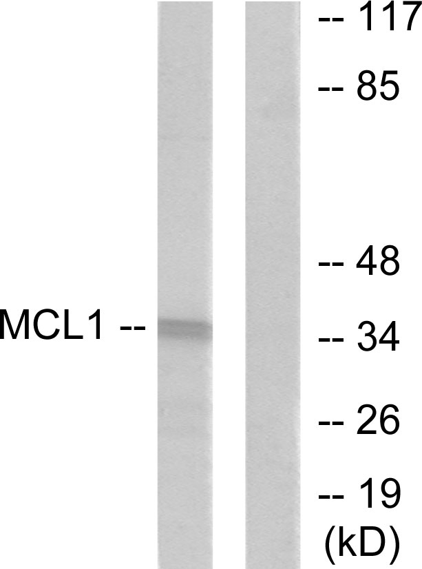
Figure 1. Western blot analysis of MCL1 using anti-MCL1 antibody (PB9132). Electrophoresis was performed on a 5-20% SDS-PAGE gel at 70V (Stacking gel) / 90V (Resolving gel) for 2-3 hours. Lane 1: recombinant human MCL1 protein 0.5 ng. After electrophoresis, proteins were transferred to a nitrocellulose membrane at 150 mA for 50-90 minutes. Blocked the membrane with 5% non-fat milk/TBS for 1.5 hour at RT. The membrane was incubated with rabbit anti-MCL1 antigen affinity purified polyclonal antibody (Catalog # PB9132) at 0.5 microg/mL overnight at 4°C, then washed with TBS-0.1%Tween 3 times with 5 minutes each and probed with a goat anti-rabbit IgG-HRP secondary antibody at a dilution of 1:5000 for 1.5 hour at RT. The signal is developed using an Enhanced Chemiluminescent detection (ECL) kit (Catalog # EK1002) with Tanon 5200 system. A specific band was detected for MCL1 at approximately 40 kDa. The expected band size for MCL1 is at 40 kDa.
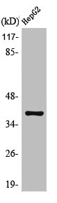
. Electrophoresis was performed on a 5-20% SDS-PAGE gel at 70V (Stacking gel) / 90V (Resolving gel) for 2-3 hours. The sample well of each lane was loaded with 30 ug of sample under reducing conditions. Lane 1: rat spleen tissue lysates, Lane 2: human HepG2 whole cell lysates, Lane 3: human MCF-7 whole cell lysates, Lane 4: human COLO320 whole cell lysates. After electrophoresis, proteins were transferred to a nitrocellulose membrane at 150 mA for 50-90 minutes. Blocked the membrane with 5% non-fat milk/TBS for 1.5 hour at RT. The membrane was incubated with rabbit anti-MCL1 antigen affinity purified polyclonal antibody (Catalog # PB9132) at 0.5 microg/mL overnight at 4°C, then washed with TBS-0.1%Tween 3 times with 5 minutes each and probed with a goat anti-rabbit IgG-HRP secondary antibody at a dilution of 1:5000 for 1.5 hour at RT. The signal is developed using an Enhanced Chemiluminescent detection (ECL) kit (Catalog # EK1002) with Tanon 5200 system. A specific band was detected for MCL1 at approximately 37 kDa. The expected band size for MCL1 is at 37 kDa.")
. MCL1 was detected in a paraffin-embedded section of rat intestine tissue. Heat mediated antigen retrieval was performed in EDTA buffer (pH 8.0, epitope retrieval solution). The tissue section was blocked with 10% goat serum. The tissue section was then incubated with 1 microg/ml rabbit anti-MCL1 Antibody (PB9132) overnight at 4°C. Biotinylated goat anti-rabbit IgG was used as secondary antibody and incubated for 30 minutes at 37°C. The tissue section was developed using Strepavidin-Biotin-Complex (SABC) (Catalog # SA1022) with DAB as the chromogen.")
. MCL1 was detected in a paraffin-embedded section of human intestinal cancer tissue. Heat mediated antigen retrieval was performed in EDTA buffer (pH 8.0, epitope retrieval solution). The tissue section was blocked with 10% goat serum. The tissue section was then incubated with 1 microg/ml rabbit anti-MCL1 Antibody (PB9132) overnight at 4°C. Biotinylated goat anti-rabbit IgG was used as secondary antibody and incubated for 30 minutes at 37°C. The tissue section was developed using Strepavidin-Biotin-Complex (SABC) (Catalog # SA1022) with DAB as the chromogen.")
. MCL1 was detected in a paraffin-embedded section of mouse intestine tissue. Heat mediated antigen retrieval was performed in EDTA buffer (pH 8.0, epitope retrieval solution). The tissue section was blocked with 10% goat serum. The tissue section was then incubated with 1 microg/ml rabbit anti-MCL1 Antibody (PB9132) overnight at 4°C. Biotinylated goat anti-rabbit IgG was used as secondary antibody and incubated for 30 minutes at 37°C. The tissue section was developed using Strepavidin-Biotin-Complex (SABC) (Catalog # SA1022) with DAB as the chromogen.")
Figure 1. Western blot analysis of MCL1 using anti-MCL1 antibody (PB9132). Electrophoresis was performed on a 5-20% SDS-PAGE gel at 70V (Stacking gel) / 90V (Resolving gel) for 2-3 hours. Lane 1: recombinant human MCL1 protein 0.5 ng. After electrophoresis, proteins were transferred to a nitrocellulose membrane at 150 mA for 50-90 minutes. Blocked the membrane with 5% non-fat milk/TBS for 1.5 hour at RT. The membrane was incubated with rabbit anti-MCL1 antigen affinity purified polyclonal antibody (Catalog # PB9132) at 0.5 microg/mL overnight at 4°C, then washed with TBS-0.1%Tween 3 times with 5 minutes each and probed with a goat anti-rabbit IgG-HRP secondary antibody at a dilution of 1:5000 for 1.5 hour at RT. The signal is developed using an Enhanced Chemiluminescent detection (ECL) kit (Catalog # EK1002) with Tanon 5200 system. A specific band was detected for MCL1 at approximately 40 kDa. The expected band size for MCL1 is at 40 kDa.
Anti-MCL1 Antibody Picoband(r)
PB9132-DYLIGHT488
ApplicationsWestern Blot, ImmunoHistoChemistry
Product group Antibodies
ReactivityHuman, Mouse, Rat
TargetMCL1
Overview
- SupplierBoster Bio
- Product NameAnti-MCL1 Antibody Picoband(r)
- Delivery Days Customer9
- Application Supplier NoteWB: The detection limit for MCL1 is approximately 0.25ng/lane under reducing conditions. Tested Species: In-house tested species with positive results. By Heat: Boiling the paraffin sections in 10mM citrate buffer, pH6.0, for 20mins is required for the staining of formalin/paraffin sections. Other applications have not been tested. Optimal dilutions should be determined by end users.
- ApplicationsWestern Blot, ImmunoHistoChemistry
- CertificationResearch Use Only
- ClonalityPolyclonal
- Concentration500 ug/ml
- ConjugateDyLight 488
- Gene ID4170
- Target nameMCL1
- Target descriptionMCL1 apoptosis regulator, BCL2 family member
- Target synonymsBCL2L3, EAT, MCL1-ES, MCL1L, MCL1S, Mcl-1, TM, bcl2-L-3, mcl1/EAT, induced myeloid leukemia cell differentiation protein Mcl-1, BCL2 family apoptosis regulator, MCL1, BCL2 family apoptosis regulator, bcl-2-like protein 3, bcl-2-related protein EAT/mcl1, myeloid cell leukemia 1, myeloid cell leukemia ES, myeloid cell leukemia sequence 1 (BCL2-related)
- HostRabbit
- IsotypeIgG
- Protein IDQ07820
- Protein NameInduced myeloid leukemia cell differentiation protein Mcl-1
- Scientific DescriptionBoster Bio Anti-MCL1 Antibody Picoband® catalog # PB9132. Tested in IHC, WB applications. This antibody reacts with Human, Mouse, Rat. The brand Picoband indicates this is a premium antibody that guarantees superior quality, high affinity, and strong signals with minimal background in Western blot applications. Only our best-performing antibodies are designated as Picoband, ensuring unmatched performance.
- ReactivityHuman, Mouse, Rat
- Storage Instruction-20°C,2°C to 8°C
- UNSPSC12352203
Related products

Product group Antibodies
Anti-MCL1 Antibody Picoband(r)A00712-2-CARRIER-FREE
ApplicationsFlow Cytometry, ImmunoFluorescence, Western Blot, ELISA, ImmunoCytoChemistry
ReactivityHuman, Mouse, Rat
TargetMCL1
- SizePrice
Product group Antibodies
MCL1 Polyclonal AntibodyCAC14605
ApplicationsImmunoFluorescence, Western Blot, ELISA, ImmunoHistoChemistry
TargetMCL1
- SizePrice
Product group Antibodies
Anti-MCL1 Antibody144-61582
ApplicationsImmunoFluorescence, Western Blot, ImmunoHistoChemistry
ReactivityHuman, Mouse
TargetMCL1
- SizePrice
Product group Antibodies

Goat anti-MCL1 AntibodyEB06380
ApplicationsWestern Blot, ELISA
ReactivityBovine, Canine, Human, Mouse, Porcine, Rat
TargetMCL1
- SizePrice
Product group Antibodies
References
MCL1 antibodyGTX102026
ApplicationsImmunoFluorescence, Western Blot, ImmunoCytoChemistry, ImmunoHistoChemistry, ImmunoHistoChemistry Paraffin
ReactivityHuman, Mouse
TargetMCL1
- SizePrice

Product group Antibodies
Anti-MCL1 AntibodyAMAB90859
ApplicationsWestern Blot, ImmunoCytoChemistry, ImmunoHistoChemistry
ReactivityHuman
TargetMCL1
- SizePrice
Product group Antibodies
Anti-MCL1 AntibodyA96164
ApplicationsWestern Blot, ELISA, ImmunoHistoChemistry
ReactivityHuman, Mouse, Rat
- SizePrice
Product group Antibodies
MCL1 AntibodyCSB-PA003207
ApplicationsWestern Blot, ELISA, ImmunoHistoChemistry
ReactivityHuman, Mouse, Rat
TargetMCL1
- SizePrice