

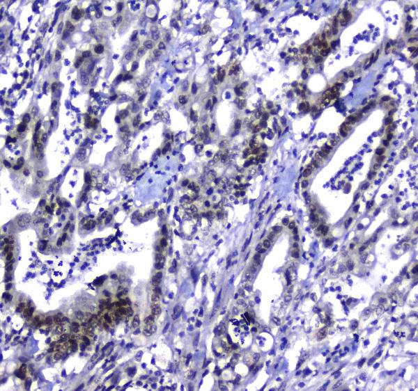
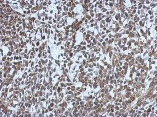
Figure 1. IHC analysis of MDC1 using anti-MDC1 antibody (A01252-2). MDC1 was detected in paraffin-embedded section of human colon cancer tissue. Heat mediated antigen retrieval was performed in citrate buffer (pH6, epitope retrieval solution) for 20 mins. The tissue section was blocked with 10% goat serum. The tissue section was then incubated with 1microg/ml rabbit anti-MDC1 Antibody (A01252-2) overnight at 4°C. Biotinylated goat anti-rabbit IgG was used as secondary antibody and incubated for 30 minutes at 37°C. The tissue section was developed using Strepavidin-Biotin-Complex (SABC)(Catalog # SA1022) with DAB as the chromogen.
. MDC1 was detected in paraffin-embedded section of human lung cancer tissue. Heat mediated antigen retrieval was performed in citrate buffer (pH6, epitope retrieval solution) for 20 mins. The tissue section was blocked with 10% goat serum. The tissue section was then incubated with 1microg/ml rabbit anti-MDC1 Antibody (A01252-2) overnight at 4°C. Biotinylated goat anti-rabbit IgG was used as secondary antibody and incubated for 30 minutes at 37°C. The tissue section was developed using Strepavidin-Biotin-Complex (SABC)(Catalog # SA1022) with DAB as the chromogen.")
. MDC1 was detected in paraffin-embedded section of human testis tissue. Heat mediated antigen retrieval was performed in citrate buffer (pH6, epitope retrieval solution) for 20 mins. The tissue section was blocked with 10% goat serum. The tissue section was then incubated with 1microg/ml rabbit anti-MDC1 Antibody (A01252-2) overnight at 4°C. Biotinylated goat anti-rabbit IgG was used as secondary antibody and incubated for 30 minutes at 37°C. The tissue section was developed using Strepavidin-Biotin-Complex (SABC)(Catalog # SA1022) with DAB as the chromogen.")
. MDC1 was detected in immunocytochemical section of A431 cells. Enzyme antigen retrieval was performed using IHC enzyme antigen retrieval reagent (AR0022) for 15 mins. The cells were blocked with 10% goat serum. And then incubated with 2microg/mL rabbit anti-MDC1 Antibody (A01252-2) overnight at 4°C. DyLight®488 Conjugated Goat Anti-Rabbit IgG (BA1127) was used as secondary antibody at 1:100 dilution and incubated for 30 minutes at 37°C. The section was counterstained with DAPI. Visualize using a fluorescence microscope and filter sets appropriate for the label used.")
. Overlay histogram showing 293T cells stained with A01252-2 (Blue line). To facilitate intracellular staining, cells were fixed with 4% paraformaldehyde and permeabilized with permeabilization buffer. The cells were blocked with 10% normal goat serum. And then incubated with rabbit anti-MDC1 Antibody (A01252-2,1microg/1x106 cells) for 30 min at 20°C. DyLight®488 conjugated goat anti-rabbit IgG (BA1127, 5-10microg/1x106 cells) was used as secondary antibody for 30 minutes at 20°C. Isotype control antibody (Green line) was rabbit IgG (1microg/1x106) used under the same conditions. Unlabelled sample without incubation with primary antibody and secondary antibody (Red line) was used as a blank control.")
Figure 1. IHC analysis of MDC1 using anti-MDC1 antibody (A01252-2). MDC1 was detected in paraffin-embedded section of human colon cancer tissue. Heat mediated antigen retrieval was performed in citrate buffer (pH6, epitope retrieval solution) for 20 mins. The tissue section was blocked with 10% goat serum. The tissue section was then incubated with 1microg/ml rabbit anti-MDC1 Antibody (A01252-2) overnight at 4°C. Biotinylated goat anti-rabbit IgG was used as secondary antibody and incubated for 30 minutes at 37°C. The tissue section was developed using Strepavidin-Biotin-Complex (SABC)(Catalog # SA1022) with DAB as the chromogen.
Anti-MDC1 Antibody
A01252-2-CARRIER-FREE
ApplicationsFlow Cytometry, ImmunoFluorescence, ELISA, ImmunoCytoChemistry, ImmunoHistoChemistry
Product group Antibodies
ReactivityHuman
TargetMDC1
Overview
- SupplierBoster Bio
- Product NameAnti-MDC1 Antibody
- Delivery Days Customer9
- ApplicationsFlow Cytometry, ImmunoFluorescence, ELISA, ImmunoCytoChemistry, ImmunoHistoChemistry
- CertificationResearch Use Only
- ClonalityPolyclonal
- Concentration500 ug/ml
- Gene ID9656
- Target nameMDC1
- Target descriptionmediator of DNA damage checkpoint 1
- Target synonymsNFBD1, mediator of DNA damage checkpoint protein 1, homologue to Drosophila photoreceptor protein calphotin, nuclear factor with BRCT domains 1
- HostRabbit
- IsotypeIgG
- Protein IDQ14676
- Protein NameMediator of DNA damage checkpoint protein 1
- Scientific DescriptionBoster Bio Anti-MDC1 Antibody Picoband® catalog # A01252-2. Tested in ELISA, Flow Cytometry, IF, IHC, ICC applications. This antibody reacts with Human.
- ReactivityHuman
- Storage Instruction-20°C,2°C to 8°C
- UNSPSC12352203
Related products
Product group Antibodies
Phospho-MDC1 (S513) AntibodyCSB-PA080045
ApplicationsELISA, ImmunoHistoChemistry
ReactivityHuman
TargetMDC1
- SizePrice
Product group Antibodies
Anti-MDC1 AntibodyA10402
ApplicationsImmunoFluorescence, Western Blot, ImmunoCytoChemistry, ImmunoHistoChemistry
ReactivityHuman
- SizePrice
Product group Antibodies
Anti-MDC1 Antibody144-66595
ApplicationsImmunoFluorescence, Western Blot, ImmunoHistoChemistry
ReactivityHuman
TargetMDC1
- SizePrice

Product group Antibodies
Anti-MDC1 AntibodyHPA006915
ApplicationsImmunoCytoChemistry, ImmunoHistoChemistry
ReactivityHuman
TargetMDC1
- SizePrice
Product group Antibodies

MDC1 AntibodyLS-C402706
ApplicationsELISA, ImmunoHistoChemistry
ReactivityHuman
TargetMDC1
- SizePrice
Product group Antibodies
MDC1 antibody [N2N3]GTX102673
ApplicationsImmunoFluorescence, Western Blot, ImmunoCytoChemistry, ImmunoHistoChemistry, ImmunoHistoChemistry Paraffin
ReactivityHuman
TargetMDC1
- SizePrice
Product group Antibodies
References
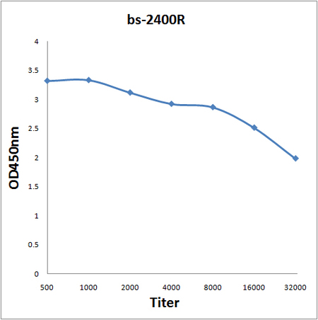
MDC1 Polyclonal AntibodyBS-2400R
ApplicationsImmunoFluorescence, Western Blot, ELISA, ImmunoCytoChemistry, ImmunoHistoChemistry, ImmunoHistoChemistry Frozen, ImmunoHistoChemistry Paraffin
ReactivityBovine, Canine, Equine, Human, Mouse, Porcine, Rat
TargetMDC1
- SizePrice