


Figure 1. Western blot analysis of MEF2C using anti-MEF2C antibody (A01131-1). Electrophoresis was performed on a 5-20% SDS-PAGE gel at 70V (Stacking gel) / 90V (Resolving gel) for 2-3 hours. The sample well of each lane was loaded with 50ug of sample under reducing conditions. Lane 1: human K562 whole cell lysates, Lane 2: human COLO320 whole cell lysates, Lane 3: human DAUDI whole cell lysates, Lane 4: human U937 whole cell lysates, Lane 5: rat brain tissue lysates. After Electrophoresis, proteins were transferred to a Nitrocellulose membrane at 150mA for 50-90 minutes. Blocked the membrane with 5% Non-fat Milk/ TBS for 1.5 hour at RT. The membrane was incubated with rabbit anti-MEF2C antigen affinity purified polyclonal antibody (Catalog # A01131-1) at 0.5 microg/mL overnight at 4°C, then washed with TBS-0.1%Tween 3 times with 5 minutes each and probed with a goat anti-rabbit IgG-HRP secondary antibody at a dilution of 1:5000 for 1.5 hour at RT. The signal is developed using an Enhanced Chemiluminescent detection (ECL) kit (Catalog # EK1002) with Tanon 5200 system. A specific band was detected for MEF2C at approximately 50-65KD. The expected band size for MEF2C is at 50-65KD.
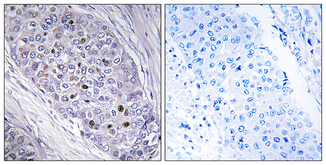
. MEF2C was detected in paraffin-embedded section of human tonsil tissue. Heat mediated antigen retrieval was performed in EDTA buffer (pH8.0, epitope retrieval solution). The tissue section was blocked with 10% goat serum. The tissue section was then incubated with 2microg/ml rabbit anti-MEF2C Antibody (A01131-1) overnight at 4°C. Biotinylated goat anti-rabbit IgG was used as secondary antibody and incubated for 30 minutes at 37°C. The tissue section was developed using Strepavidin-Biotin-Complex (SABC) (Catalog # SA1022) with DAB as the chromogen.")
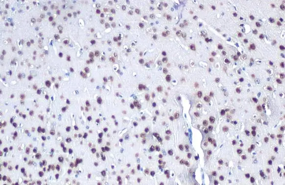
. MEF2C was detected in paraffin-embedded section of rat brain tissue. Heat mediated antigen retrieval was performed in EDTA buffer (pH8.0, epitope retrieval solution). The tissue section was blocked with 10% goat serum. The tissue section was then incubated with 2microg/ml rabbit anti-MEF2C Antibody (A01131-1) overnight at 4°C. Biotinylated goat anti-rabbit IgG was used as secondary antibody and incubated for 30 minutes at 37°C. The tissue section was developed using Strepavidin-Biotin-Complex (SABC) (Catalog # SA1022) with DAB as the chromogen.")
. MEF2C was detected in paraffin-embedded section of mouse brain tissue. Heat mediated antigen retrieval was performed in EDTA buffer (pH8.0, epitope retrieval solution). The tissue section was blocked with 10% goat serum. The tissue section was then incubated with 2microg/ml rabbit anti-MEF2C Antibody (A01131-1) overnight at 4°C. Biotinylated goat anti-rabbit IgG was used as secondary antibody and incubated for 30 minutes at 37°C. The tissue section was developed using Strepavidin-Biotin-Complex (SABC) (Catalog # SA1022) with DAB as the chromogen.")
. Overlay histogram showing HELA cells stained with A01131-1 (Blue line). To facilitate intracellular staining, cells were fixed with 4% paraformaldehyde and permeabilized with permeabilization buffer. The cells were blocked with 10% normal goat serum. And then incubated with rabbit anti-MEF2C Antibody (A01131-1, 1microg/1x106 cells) for 30 min at 20°C. DyLight®488 conjugated goat anti-rabbit IgG (BA1127, 5-10microg/1x106 cells) was used as secondary antibody for 30 minutes at 20°C. Isotype control antibody (Green line) was rabbit IgG (1microg/1x106) used under the same conditions. Unlabelled sample without incubation with primary antibody and secondary antibody (Red line) was used as a blank control.")
Figure 1. Western blot analysis of MEF2C using anti-MEF2C antibody (A01131-1). Electrophoresis was performed on a 5-20% SDS-PAGE gel at 70V (Stacking gel) / 90V (Resolving gel) for 2-3 hours. The sample well of each lane was loaded with 50ug of sample under reducing conditions. Lane 1: human K562 whole cell lysates, Lane 2: human COLO320 whole cell lysates, Lane 3: human DAUDI whole cell lysates, Lane 4: human U937 whole cell lysates, Lane 5: rat brain tissue lysates. After Electrophoresis, proteins were transferred to a Nitrocellulose membrane at 150mA for 50-90 minutes. Blocked the membrane with 5% Non-fat Milk/ TBS for 1.5 hour at RT. The membrane was incubated with rabbit anti-MEF2C antigen affinity purified polyclonal antibody (Catalog # A01131-1) at 0.5 microg/mL overnight at 4°C, then washed with TBS-0.1%Tween 3 times with 5 minutes each and probed with a goat anti-rabbit IgG-HRP secondary antibody at a dilution of 1:5000 for 1.5 hour at RT. The signal is developed using an Enhanced Chemiluminescent detection (ECL) kit (Catalog # EK1002) with Tanon 5200 system. A specific band was detected for MEF2C at approximately 50-65KD. The expected band size for MEF2C is at 50-65KD.
Anti-MEF2C Antibody Picoband(r)
A01131-1-CARRIER-FREE
ApplicationsFlow Cytometry, Western Blot, ImmunoHistoChemistry
Product group Antibodies
ReactivityHuman, Mouse, Rat
TargetMEF2C
Overview
- SupplierBoster Bio
- Product NameAnti-MEF2C Antibody Picoband(r)
- Delivery Days Customer9
- ApplicationsFlow Cytometry, Western Blot, ImmunoHistoChemistry
- CertificationResearch Use Only
- ClonalityPolyclonal
- Concentration500 ug/ml
- Gene ID4208
- Target nameMEF2C
- Target descriptionmyocyte enhancer factor 2C
- Target synonymsC5DELq14.3, DEL5q14.3, NEDHSIL, myocyte-specific enhancer factor 2C, MADS box transcription enhancer factor 2, polypeptide C
- HostRabbit
- IsotypeIgG
- Protein IDQ06413
- Protein NameMyocyte-specific enhancer factor 2C
- Scientific DescriptionBoster Bio Anti-MEF2C Antibody Picoband® catalog # A01131-1. Tested in Flow Cytometry, IHC, WB applications. This antibody reacts with Human, Mouse, Rat. The brand Picoband indicates this is a premium antibody that guarantees superior quality, high affinity, and strong signals with minimal background in Western blot applications. Only our best-performing antibodies are designated as Picoband, ensuring unmatched performance.
- ReactivityHuman, Mouse, Rat
- Storage Instruction-20°C,2°C to 8°C
- UNSPSC12352203
Related products
Product group Antibodies
Anti-MEF2C AntibodyA98862
ApplicationsELISA, ImmunoHistoChemistry
ReactivityHuman, Mouse
- SizePrice
Product group Antibodies

MEF2C (Phospho-Ser396) AntibodyABX012699
ApplicationsWestern Blot, ELISA, ImmunoHistoChemistry
- SizePrice
Product group Antibodies

Anti-MFE2C [AbAb-MFE2C]AB04171-1.1
ApplicationsImmunoFluorescence, Western Blot, ELISA, Other Application
ReactivityHuman
TargetMEF2C
- SizePrice

Product group Antibodies
Anti-MEF2C AntibodyAMAB90727
ApplicationsWestern Blot, ImmunoCytoChemistry, ImmunoHistoChemistry
ReactivityHuman
TargetMEF2C
- SizePrice
Product group Antibodies
ApplicationsWestern Blot, ELISA
ReactivityCanine, Human, Mouse, Porcine, Rabbit, Rat
TargetMEF2C
- SizePrice
Product group Antibodies
Phospho-MEF2C (S396) AntibodyCSB-PA010169
ApplicationsWestern Blot, ELISA, ImmunoHistoChemistry
ReactivityHuman, Mouse
TargetMEF2C
- SizePrice
Product group Antibodies
MEF2C antibodyGTX105433
ApplicationsImmunoFluorescence, ImmunoPrecipitation, Western Blot, ImmunoCytoChemistry, ImmunoHistoChemistry, ImmunoHistoChemistry Frozen, ImmunoHistoChemistry Paraffin
ReactivityHuman, Mouse
TargetMEF2C
- SizePrice
Product group Antibodies

MEF2C AntibodyLS-C763475
ApplicationsImmunoHistoChemistry
ReactivityHuman
TargetMEF2C
- SizePrice