


Immunofluorescent staining of human cell line SiHa shows localization to nucleus & nucleoli.
Immunofluorescent staining of human cell line SiHa shows localization to nucleus & nucleoli.
Anti-MEIOB Antibody
HPA042547
ApplicationsImmunoCytoChemistry
Product group Antibodies
ReactivityHuman
TargetMEIOB
Overview
- SupplierAtlas Antibodies
- Product NameAnti-MEIOB Antibody
- Delivery Days Customer4
- ApplicationsImmunoCytoChemistry
- CertificationResearch Use Only
- ClonalityPolyclonal
- ConjugateUnconjugated
- Gene ID254528
- Target nameMEIOB
- Target descriptionmeiosis specific with OB-fold
- Target synonymsC16orf73, POF23, SPGF22, gs129, meiosis-specific with OB domain-containing protein, meiosis specific with OB domains
- HostRabbit
- IsotypeIgG
- Protein IDQ8N635
- Protein NameMeiosis-specific with OB domain-containing protein
- Scientific DescriptionRecombinant Protein Epitope Signature Tag (PrEST) antigen sequence
- ReactivityHuman
- Storage Instruction-20°C,2°C to 8°C
- UNSPSC41116161
Datasheet
MSDS
Related products

Product group Antibodies
Anti-MEIOB Antibody Picoband(r)A13211-2-CARRIER-FREE
ApplicationsFlow Cytometry, ImmunoFluorescence, Western Blot, ELISA, ImmunoCytoChemistry
ReactivityHuman, Mouse, Rat
TargetMEIOB
- SizePrice
Product group Antibodies
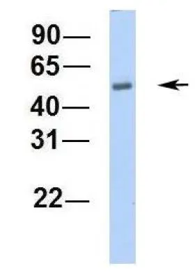
C16orf73 antibody, InternalGTX45259
ApplicationsWestern Blot
ReactivityHuman
TargetMEIOB
- SizePrice
Product group Antibodies

C16orf73 Antibody (C-Terminus)LS-C116620
ApplicationsWestern Blot
ReactivityHuman
TargetMEIOB
- SizePrice