

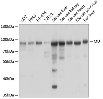


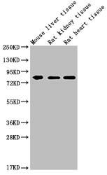
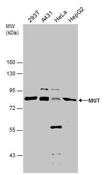
Anti-Methylmalonyl Coenzyme A mutase Antibody
A14496
ApplicationsImmunoFluorescence, Western Blot, ImmunoCytoChemistry
Product group Antibodies
ReactivityHuman, Mouse, Rat
Overview
- SupplierAntibodies.com
- Product NameAnti-Methylmalonyl Coenzyme A mutase Antibody
- Delivery Days Customer7
- ApplicationsImmunoFluorescence, Western Blot, ImmunoCytoChemistry
- CertificationResearch Use Only
- ClonalityPolyclonal
- ConjugateUnconjugated
- HostRabbit
- IsotypeIgG
- Scientific DescriptionRabbit polyclonal antibody to Methylmalonyl Coenzyme A mutase.
- ReactivityHuman, Mouse, Rat
- UNSPSC12352203
Related products

Product group Antibodies
Anti-Methylmalonyl Coenzyme A mutase Antibody Picoband(r)A01065-CARRIER-FREE
ApplicationsFlow Cytometry, ImmunoFluorescence, Western Blot, ImmunoCytoChemistry, ImmunoHistoChemistry
ReactivityHuman, Mouse, Rat
TargetMMUT
- SizePrice
Product group Antibodies
Anti-MUT Antibody144-03969
ApplicationsWestern Blot
ReactivityHuman, Mouse, Rat
TargetMMUT
- SizePrice
Product group Antibodies

MUT / MCM AntibodyLS-C667687
ApplicationsWestern Blot
ReactivityHuman
TargetMMUT
- SizePrice
Product group Antibodies
ApplicationsWestern Blot, ImmunoCytoChemistry
ReactivityHuman, Mouse, Rat
TargetMMUT
- SizePrice
Product group Antibodies
MMUT Monoclonal AntibodyCSB-MA015243A0M
ApplicationsWestern Blot, ELISA
ReactivityHuman, Mouse, Rat
TargetMMUT
- SizePrice
Product group Antibodies
MMUT Monoclonal AntibodyCAC13712
ApplicationsWestern Blot, ELISA
ReactivityMouse, Rat
TargetMMUT
- SizePrice
Product group Antibodies
MUT antibodyGTX130666
ApplicationsWestern Blot
ReactivityHuman
TargetMMUT
- SizePrice

Product group Antibodies
Anti-MUT AntibodyHPA035971
ApplicationsWestern Blot, ImmunoHistoChemistry
ReactivityHuman, Mouse, Rat
TargetMMUT
- SizePrice
Product group Antibodies
TargetMMUT
- SizePrice