


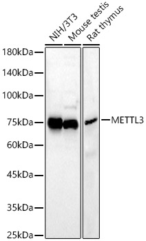
Figure 1. Western blot analysis of METTL3 using anti-METTL3 antibody (A01758-1). Electrophoresis was performed on a 5-20% SDS-PAGE gel at 70V (Stacking gel) / 90V (Resolving gel) for 2-3 hours. The sample well of each lane was loaded with 30 ug of sample under reducing conditions. Lane 1: human HepG2 whole cell lysates, Lane 2: human PC-3 whole cell lysates, Lane 3: human A549 whole cell lysates, Lane 4: human Hacat whole cell lysates, Lane 5: rat brain tissue lysates, Lane 6: rat testis tissue lysates, Lane 7: mouse brain tissue lysates. After electrophoresis, proteins were transferred to a nitrocellulose membrane at 150 mA for 50-90 minutes. Blocked the membrane with 5% non-fat milk/TBS for 1.5 hour at RT. The membrane was incubated with rabbit anti-METTL3 antigen affinity purified polyclonal antibody (Catalog # A01758-1) at 0.5 microg/mL overnight at 4°C, then washed with TBS-0.1%Tween 3 times with 5 minutes each and probed with a goat anti-rabbit IgG-HRP secondary antibody at a dilution of 1:5000 for 1.5 hour at RT. The signal is developed using an Enhanced Chemiluminescent detection (ECL) kit (Catalog # EK1002) with Tanon 5200 system. A specific band was detected for METTL3 at approximately 64 kDa. The expected band size for METTL3 is at 64 kDa.
. Overlay histogram showing HEPA1-6 cells stained with A01758-1 (Blue line). To facilitate intracellular staining, cells were fixed with 4% paraformaldehyde and permeabilized with permeabilization buffer. The cells were blocked with 10% normal goat serum. And then incubated with rabbit anti-METTL3 Antibody (A01758-1, 1 microg/1x106 cells) for 30 min at 20°C. DyLight®488 conjugated goat anti-rabbit IgG (BA1127, 5-10 microg/1x106 cells) was used as secondary antibody for 30 minutes at 20°C. Isotype control antibody (Green line) was rabbit IgG (1 microg/1x106) used under the same conditions. Unlabelled sample without incubation with primary antibody and secondary antibody (Red line) was used as a blank control.")
Figure 1. Western blot analysis of METTL3 using anti-METTL3 antibody (A01758-1). Electrophoresis was performed on a 5-20% SDS-PAGE gel at 70V (Stacking gel) / 90V (Resolving gel) for 2-3 hours. The sample well of each lane was loaded with 30 ug of sample under reducing conditions. Lane 1: human HepG2 whole cell lysates, Lane 2: human PC-3 whole cell lysates, Lane 3: human A549 whole cell lysates, Lane 4: human Hacat whole cell lysates, Lane 5: rat brain tissue lysates, Lane 6: rat testis tissue lysates, Lane 7: mouse brain tissue lysates. After electrophoresis, proteins were transferred to a nitrocellulose membrane at 150 mA for 50-90 minutes. Blocked the membrane with 5% non-fat milk/TBS for 1.5 hour at RT. The membrane was incubated with rabbit anti-METTL3 antigen affinity purified polyclonal antibody (Catalog # A01758-1) at 0.5 microg/mL overnight at 4°C, then washed with TBS-0.1%Tween 3 times with 5 minutes each and probed with a goat anti-rabbit IgG-HRP secondary antibody at a dilution of 1:5000 for 1.5 hour at RT. The signal is developed using an Enhanced Chemiluminescent detection (ECL) kit (Catalog # EK1002) with Tanon 5200 system. A specific band was detected for METTL3 at approximately 64 kDa. The expected band size for METTL3 is at 64 kDa.
Anti-METTL3 Antibody Picoband(r)
A01758-1-CARRIER-FREE
ApplicationsFlow Cytometry, Western Blot, ELISA
Product group Antibodies
ReactivityHuman, Mouse, Rat
TargetMETTL3
Overview
- SupplierBoster Bio
- Product NameAnti-METTL3 Antibody Picoband(r)
- Delivery Days Customer9
- ApplicationsFlow Cytometry, Western Blot, ELISA
- CertificationResearch Use Only
- ClonalityPolyclonal
- Concentration500 ug/ml
- Gene ID56339
- Target nameMETTL3
- Target descriptionmethyltransferase 3, N6-adenosine-methyltransferase complex catalytic subunit
- Target synonymsIME4, M6A, MT-A70, Spo8, hMETTL3, N(6)-adenosine-methyltransferase catalytic subunit METTL3, N(6)-adenosine-methyltransferase 70 kDa subunit, N6-adenosine-methyltransferase 70 kDa subunit, N6-adenosine-methyltransferase catalytic subunit, adoMet-binding subunit of the human mRNA (N6-adenosine)-methyltransferase, mRNA (2'-O-methyladenosine-N(6)-)-methyltransferase, mRNA m(6)A methyltransferase, methyltransferase like 3, methyltransferase-like protein 3
- HostRabbit
- IsotypeIgG
- Protein IDQ86U44
- Protein NameN(6)-adenosine-methyltransferase catalytic subunit METTL3
- Scientific DescriptionBoster Bio Anti-METTL3/METTL3 Antibody Picoband® catalog # A01758-1. Tested in ELISA, Flow Cytometry, WB applications. This antibody reacts with Human, Mouse, Rat. The brand Picoband indicates this is a premium antibody that guarantees superior quality, high affinity, and strong signals with minimal background in Western blot applications. Only our best-performing antibodies are designated as Picoband, ensuring unmatched performance.
- ReactivityHuman, Mouse, Rat
- Storage Instruction-20°C,2°C to 8°C
- UNSPSC12352203
Related products
Product group Antibodies
Anti-METTL3 AntibodyA12244
ApplicationsImmunoFluorescence, Western Blot, ImmunoCytoChemistry, ImmunoHistoChemistry
ReactivityHuman, Mouse, Rat
- SizePrice
Product group Antibodies
Anti-METTL3 Antibody144-08370
ApplicationsImmunoFluorescence, ImmunoPrecipitation, Western Blot, ImmunoHistoChemistry
ReactivityHuman, Mouse, Rat
TargetMETTL3
- SizePrice
Product group Antibodies
METTL3 Polyclonal AntibodyBS-17609R
ApplicationsImmunoFluorescence, Western Blot, ELISA, ImmunoCytoChemistry, ImmunoHistoChemistry, ImmunoHistoChemistry Frozen, ImmunoHistoChemistry Paraffin
ReactivityBovine, Human, Mouse, Porcine, Rabbit, Rat, Sheep
TargetMETTL3
- SizePrice
Product group Antibodies
METTL3 AntibodyCSB-PA013732GA01HU
ApplicationsWestern Blot, ELISA
ReactivityHuman, Mouse, Rat
TargetMETTL3
- SizePrice
Product group Antibodies

METTL3 AntibodyLS-C409906
ApplicationsImmunoPrecipitation, Western Blot, ImmunoHistoChemistry
ReactivityHuman, Mouse
TargetMETTL3
- SizePrice

Product group Antibodies
Anti-METTL3 AntibodyHPA079662
ApplicationsImmunoCytoChemistry
ReactivityHuman
TargetMETTL3
- SizePrice
![Various whole cell extracts (30 μg) were separated by 7.5% SDS-PAGE, and the membrane was blotted with METTL3 antibody [N2C2], Internal (GTX105037) diluted at 1:10000. The HRP-conjugated anti-rabbit IgG antibody (GTX213110-01) was used to detect the primary antibody, and the signal was developed with Trident ECL plus-Enhanced.](https://www.genetex.com/upload/website/prouct_img/normal/GTX105037/GTX105037_40044_20191227_WB_R_w_23060120_981.webp)
Product group Antibodies
METTL3 antibody [N2C2], InternalGTX105037
ApplicationsWestern Blot, ImmunoHistoChemistry, ImmunoHistoChemistry Paraffin
ReactivityHuman, Rat
TargetMETTL3
- SizePrice
Product group Antibodies
ApplicationsImmunoFluorescence, ImmunoPrecipitation, Western Blot, ELISA, ImmunoCytoChemistry
ReactivityHuman
TargetMETTL3
- SizePrice