

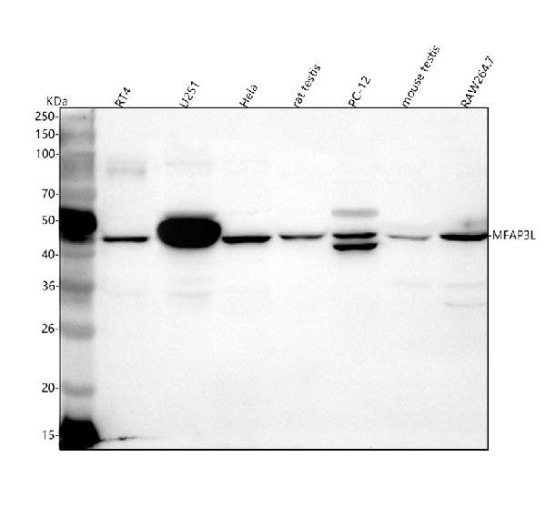
Figure 1. Western blot analysis of MFAP3L using anti-MFAP3L antibody (A12581). Electrophoresis was performed on a 5-20% SDS-PAGE gel at 70V (Stacking gel) / 90V (Resolving gel) for 2-3 hours. The sample well of each lane was loaded with 30 ug of sample under reducing conditions. Lane 1: human RT4 whole cell lysates, Lane 2: human U251 whole cell lysates, Lane 3: human Hela whole cell lysates, Lane 4: rat testis tissue lysates, Lane 5: rat PC-12 whole cell lysates, Lane 6: mouse testis tissue lysates, Lane 7: mouse RAW264.7 whole cell lysates. After electrophoresis, proteins were transferred to a nitrocellulose membrane at 150 mA for 50-90 minutes. Blocked the membrane with 5% non-fat milk/TBS for 1.5 hour at RT. The membrane was incubated with rabbit anti-MFAP3L antigen affinity purified polyclonal antibody (Catalog # A12581) at 0.5 microg/mL overnight at 4°C, then washed with TBS-0.1%Tween 3 times with 5 minutes each and probed with a goat anti-rabbit IgG-HRP secondary antibody at a dilution of 1:5000 for 1.5 hour at RT. The signal is developed using an Enhanced Chemiluminescent detection (ECL) kit (Catalog # EK1002) with Tanon 5200 system. A specific band was detected for MFAP3L at approximately 45 kDa. The expected band size for MFAP3L is at 45 kDa.
. MFAP3L was detected in an immunocytochemical section of HELA cells. Enzyme antigen retrieval was performed using IHC enzyme antigen retrieval reagent (AR0022) for 15 mins. The cells were blocked with 10% goat serum. And then incubated with 5 microg/mL rabbit anti-MFAP3L Antibody (A12581) overnight at 4°C. DyLight®488 Conjugated Goat Anti-Rabbit IgG (BA1127) was used as secondary antibody at 1:500 dilution and incubated for 30 minutes at 37°C. The section was counterstained with DAPI. Visualize using a fluorescence microscope and filter sets appropriate for the label used.")
Figure 1. Western blot analysis of MFAP3L using anti-MFAP3L antibody (A12581). Electrophoresis was performed on a 5-20% SDS-PAGE gel at 70V (Stacking gel) / 90V (Resolving gel) for 2-3 hours. The sample well of each lane was loaded with 30 ug of sample under reducing conditions. Lane 1: human RT4 whole cell lysates, Lane 2: human U251 whole cell lysates, Lane 3: human Hela whole cell lysates, Lane 4: rat testis tissue lysates, Lane 5: rat PC-12 whole cell lysates, Lane 6: mouse testis tissue lysates, Lane 7: mouse RAW264.7 whole cell lysates. After electrophoresis, proteins were transferred to a nitrocellulose membrane at 150 mA for 50-90 minutes. Blocked the membrane with 5% non-fat milk/TBS for 1.5 hour at RT. The membrane was incubated with rabbit anti-MFAP3L antigen affinity purified polyclonal antibody (Catalog # A12581) at 0.5 microg/mL overnight at 4°C, then washed with TBS-0.1%Tween 3 times with 5 minutes each and probed with a goat anti-rabbit IgG-HRP secondary antibody at a dilution of 1:5000 for 1.5 hour at RT. The signal is developed using an Enhanced Chemiluminescent detection (ECL) kit (Catalog # EK1002) with Tanon 5200 system. A specific band was detected for MFAP3L at approximately 45 kDa. The expected band size for MFAP3L is at 45 kDa.
Anti-MFAP3L Antibody Picoband(r)
A12581-CARRIER-FREE
ApplicationsImmunoFluorescence, Western Blot, ELISA, ImmunoCytoChemistry
Product group Antibodies
ReactivityHuman, Mouse, Rat
TargetMFAP3L
Overview
- SupplierBoster Bio
- Product NameAnti-MFAP3L Antibody Picoband(r)
- Delivery Days Customer9
- ApplicationsImmunoFluorescence, Western Blot, ELISA, ImmunoCytoChemistry
- CertificationResearch Use Only
- ClonalityPolyclonal
- Concentration500 ug/ml
- Gene ID9848
- Target nameMFAP3L
- Target descriptionmicrofibril associated protein 3 like
- Target synonymsNYD-sp9, microfibrillar-associated protein 3-like, microfi brillar-associated protein 3-like, microfibrillar associated protein 3 like, testis development protein NYD-SP9
- HostRabbit
- Protein IDO75121
- Protein NameMicrofibrillar-associated protein 3-like
- Scientific DescriptionBoster Bio Anti-MFAP3L Antibody Picoband® catalog # A12581. Tested in WB, ICC/IF, ELISA applications. This antibody reacts with Human, Mouse, Rat. The brand Picoband indicates this is a premium antibody that guarantees superior quality, high affinity, and strong signals with minimal background in Western blot applications. Only our best-performing antibodies are designated as Picoband, ensuring unmatched performance.
- ReactivityHuman, Mouse, Rat
- Storage Instruction-20°C,2°C to 8°C
- UNSPSC12352203
Related products

Product group Antibodies
Anti-MFAP3L AntibodyA45465
ApplicationsImmunoHistoChemistry
ReactivityHuman
- SizePrice
Product group Antibodies
MFAP3L AntibodyCSB-PA013748LA01HU
ApplicationsImmunoFluorescence, ELISA, ImmunoHistoChemistry
ReactivityHuman
TargetMFAP3L
- SizePrice
Product group Antibodies

MFAP3L / NYD-Sp9 AntibodyLS-C401838
ApplicationsELISA, ImmunoHistoChemistry
ReactivityHuman, Mouse, Rat
TargetMFAP3L
- SizePrice
Product group Antibodies
MFAP3L antibody, N-termGTX30779
ApplicationsWestern Blot
ReactivityHuman
TargetMFAP3L
- SizePrice

Product group Antibodies
Anti-MFAP3L AntibodyHPA017986
ApplicationsImmunoHistoChemistry
ReactivityHuman
TargetMFAP3L
- SizePrice