

Immunohistochemical staining of human gallbladder shows weak cytoplasmic positivity in glandular cells.
Immunohistochemical staining of human gallbladder shows weak cytoplasmic positivity in glandular cells.
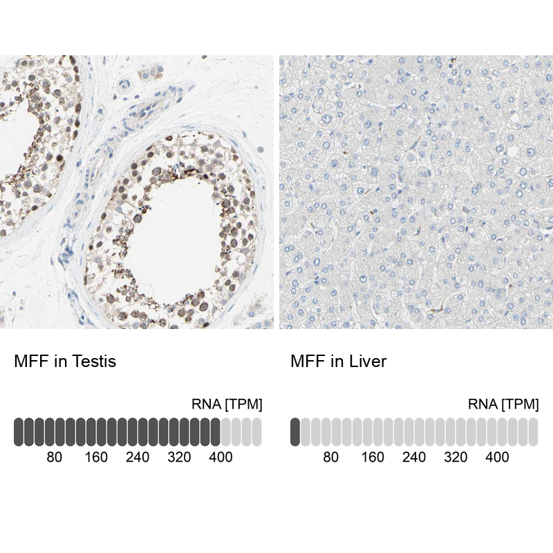
Anti-MFF Antibody
HPA010968
ApplicationsImmunoHistoChemistry
Product group Antibodies
ReactivityHuman
TargetMFF
Overview
- SupplierAtlas Antibodies
- Product NameAnti-MFF Antibody
- Delivery Days Customer4
- ApplicationsImmunoHistoChemistry
- CertificationResearch Use Only
- ClonalityPolyclonal
- ConjugateUnconjugated
- Gene ID56947
- Target nameMFF
- Target descriptionmitochondrial fission factor
- Target synonymsC2orf33, EMPF2, GL004, mitochondrial fission factor
- HostRabbit
- IsotypeIgG
- Protein IDQ9GZY8
- Protein NameMitochondrial fission factor
- Scientific DescriptionRecombinant Protein Epitope Signature Tag (PrEST) antigen sequence
- ReactivityHuman
- Storage Instruction-20°C,2°C to 8°C
- UNSPSC41116161
Datasheet
MSDS
Related products

Product group Antibodies
Anti-MFF Antibody Picoband(r)A02563-1-CARRIER-FREE
ApplicationsFlow Cytometry, ImmunoFluorescence, Western Blot, ELISA, ImmunoCytoChemistry, ImmunoHistoChemistry
ReactivityHuman, Mouse, Rat
TargetMFF
- SizePrice
Product group Antibodies
Anti-MFF Antibody144-65840
ApplicationsWestern Blot, ImmunoHistoChemistry
ReactivityHuman
TargetMFF
- SizePrice
Product group Antibodies

ApplicationsWestern Blot, ELISA
- SizePrice
Product group Antibodies
Anti-MFF AntibodyA14609
ApplicationsWestern Blot, ImmunoHistoChemistry
ReactivityHuman, Mouse, Rat
- SizePrice
Product group Antibodies

MFF AntibodyLS-C747496
ApplicationsWestern Blot
ReactivityHuman
TargetMFF
- SizePrice
Product group Antibodies
MFF Polyclonal AntibodyBS-7628R
ApplicationsImmunoFluorescence, Western Blot, ELISA, ImmunoCytoChemistry, ImmunoHistoChemistry, ImmunoHistoChemistry Frozen, ImmunoHistoChemistry Paraffin
ReactivityBovine, Chicken, Equine, Human, Mouse, Porcine, Rabbit, Rat, Sheep, Zebra Fish
- SizePrice
Product group Antibodies
MFF AntibodyCSB-PA013751LA01HU
ApplicationsWestern Blot, ELISA
ReactivityHuman
TargetMFF
- SizePrice
Product group Antibodies
MFF Polyclonal AntibodyCAC15837
ApplicationsWestern Blot, ELISA
TargetMFF
- SizePrice