


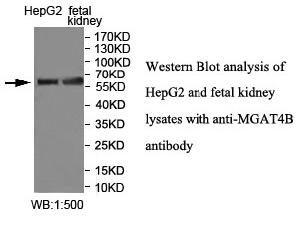
Figure 1. Western blot analysis of MGAT4B using anti-MGAT4B antibody (A10339). Electrophoresis was performed on a 5-20% SDS-PAGE gel at 70V (Stacking gel) / 90V (Resolving gel) for 2-3 hours. The sample well of each lane was loaded with 30 ug of sample under reducing conditions. Lane 1: human HepG2 whole cell lysates, Lane 2: human RT4 whole cell lysates, Lane 3: human A431 whole cell lysates, Lane 4: human Caco-2 whole cell lysates, Lane 5: rat kidney tissue lysates, Lane 6: mouse kidney tissue lysates. After electrophoresis, proteins were transferred to a nitrocellulose membrane at 150 mA for 50-90 minutes. Blocked the membrane with 5% non-fat milk/TBS for 1.5 hour at RT. The membrane was incubated with rabbit anti-MGAT4B antigen affinity purified polyclonal antibody (Catalog # A10339) at 0.5 microg/mL overnight at 4°C, then washed with TBS-0.1%Tween 3 times with 5 minutes each and probed with a goat anti-rabbit IgG-HRP secondary antibody at a dilution of 1:5000 for 1.5 hour at RT. The signal is developed using an Enhanced Chemiluminescent detection (ECL) kit (Catalog # EK1002) with Tanon 5200 system. A specific band was detected for MGAT4B at approximately 70 kDa. The expected band size for MGAT4B is at 63 kDa.
. MGAT4B was detected in a paraffin-embedded section of human glioma tissue. Heat mediated antigen retrieval was performed in EDTA buffer (pH 8.0, epitope retrieval solution). The tissue section was blocked with 10% goat serum. The tissue section was then incubated with 2 microg/ml rabbit anti-MGAT4B Antibody (A10339) overnight at 4°C. Peroxidase Conjugated Goat Anti-rabbit IgG was used as secondary antibody and incubated for 30 minutes at 37°C. The tissue section was developed using HRP Conjugated Rabbit IgG Super Vision Assay Kit (Catalog # SV0002) with DAB as the chromogen.")
Figure 1. Western blot analysis of MGAT4B using anti-MGAT4B antibody (A10339). Electrophoresis was performed on a 5-20% SDS-PAGE gel at 70V (Stacking gel) / 90V (Resolving gel) for 2-3 hours. The sample well of each lane was loaded with 30 ug of sample under reducing conditions. Lane 1: human HepG2 whole cell lysates, Lane 2: human RT4 whole cell lysates, Lane 3: human A431 whole cell lysates, Lane 4: human Caco-2 whole cell lysates, Lane 5: rat kidney tissue lysates, Lane 6: mouse kidney tissue lysates. After electrophoresis, proteins were transferred to a nitrocellulose membrane at 150 mA for 50-90 minutes. Blocked the membrane with 5% non-fat milk/TBS for 1.5 hour at RT. The membrane was incubated with rabbit anti-MGAT4B antigen affinity purified polyclonal antibody (Catalog # A10339) at 0.5 microg/mL overnight at 4°C, then washed with TBS-0.1%Tween 3 times with 5 minutes each and probed with a goat anti-rabbit IgG-HRP secondary antibody at a dilution of 1:5000 for 1.5 hour at RT. The signal is developed using an Enhanced Chemiluminescent detection (ECL) kit (Catalog # EK1002) with Tanon 5200 system. A specific band was detected for MGAT4B at approximately 70 kDa. The expected band size for MGAT4B is at 63 kDa.
Anti-MGAT4B Antibody Picoband(r)
A10339-CARRIER-FREE
ApplicationsWestern Blot, ELISA, ImmunoHistoChemistry
Product group Antibodies
ReactivityHuman, Mouse, Rat
TargetMGAT4B
Overview
- SupplierBoster Bio
- Product NameAnti-MGAT4B Antibody Picoband(r)
- Delivery Days Customer9
- Application Supplier NoteTested Species: In-house tested species with positive results. Other applications have not been tested. Optimal dilutions should be determined by end users.
- ApplicationsWestern Blot, ELISA, ImmunoHistoChemistry
- CertificationResearch Use Only
- ClonalityPolyclonal
- Concentration500 ug/ml
- Gene ID11282
- Target nameMGAT4B
- Target descriptionalpha-1,3-mannosyl-glycoprotein 4-beta-N-acetylglucosaminyltransferase B
- Target synonymsGNT-IV, GNT-IVB, alpha-1,3-mannosyl-glycoprotein 4-beta-N-acetylglucosaminyltransferase B, N-acetylglucosaminyltransferase IVb, N-glycosyl-oligosaccharide-glycoprotein N-acetylglucosaminyltransferase IVb, UDP-N-acetylglucosamine: alpha-1,3-D-mannoside beta-1,4-N-acetylglucosaminyltransferase IV, UDP-N-acetylglucosamine: alpha-1,3-D-mannoside beta-1,4-N-acetylglucosaminyltransferase IVb, alpha-1,3-mannosyl-glycoprotein beta-1,4-N-acetylglucosaminyltransferase, aminyltransferase, glcNAc-T IVb, mannosyl (alpha-1,3-)-glycoprotein beta-1,4-N-acetylglucosaminyltransferase, isoenzyme B, mannosyl (alpha-1,3-)-glycoprotein beta-1,4-N-acetylglucosaminyltransferase, isozyme B
- HostRabbit
- IsotypeIgG
- Protein IDQ9UQ53
- Protein NameAlpha-1,3-mannosyl-glycoprotein 4-beta-N-acetylglucosaminyltransferase B
- Scientific DescriptionBoster Bio Anti-MGAT4B Antibody Picoband® catalog # A10339. Tested in ELISA, IHC, WB applications. This antibody reacts with Human, Mouse, Rat. The brand Picoband indicates this is a premium antibody that guarantees superior quality, high affinity, and strong signals with minimal background in Western blot applications. Only our best-performing antibodies are designated as Picoband, ensuring unmatched performance.
- ReactivityHuman, Mouse, Rat
- Storage Instruction-20°C,2°C to 8°C
- UNSPSC12352203
Related products
Product group Antibodies
Anti-MGAT4B AntibodyA47831
ApplicationsWestern Blot, ELISA, ImmunoHistoChemistry
ReactivityHuman, Mouse, Rat
- SizePrice
Product group Antibodies

MGAT4B AntibodyLS-C830970
ApplicationsELISA, ImmunoHistoChemistry
ReactivityHuman, Mouse
TargetMGAT4B
- SizePrice

Product group Antibodies
Anti-MGAT4B AntibodyHPA012804
ApplicationsImmunoCytoChemistry
ReactivityHuman
TargetMGAT4B
- SizePrice
Product group Antibodies
MGAT4B AntibodyCSB-PA890781LA01HU
ApplicationsWestern Blot, ELISA, ImmunoHistoChemistry
ReactivityHuman
TargetMGAT4B
- SizePrice
Product group Antibodies
Mgat4B Polyclonal AntibodyCAC11570
ApplicationsWestern Blot, ELISA, ImmunoHistoChemistry
TargetMGAT4B
- SizePrice
Product group Antibodies
Anti-MGAT4BY158509
ApplicationsWestern Blot, ELISA, ImmunoHistoChemistry
ReactivityHuman
- SizePrice
Product group Antibodies
Anti-MGAT4B (N-term) Antibody102-25013
ApplicationsWestern Blot
TargetMGAT4B
- SizePrice