


Immunohistochemical staining of human colon shows strong cytoplasmic positivity in glandular cells.

Immunohistochemical staining of human colon shows strong cytoplasmic positivity in glandular cells.
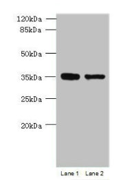
Anti-MGME1 Antibody
HPA040913
ApplicationsWestern Blot, ImmunoCytoChemistry, ImmunoHistoChemistry
Product group Antibodies
ReactivityHuman
TargetMGME1
Overview
- SupplierAtlas Antibodies
- Product NameAnti-MGME1 Antibody
- Delivery Days Customer4
- ApplicationsWestern Blot, ImmunoCytoChemistry, ImmunoHistoChemistry
- CertificationResearch Use Only
- ClonalityPolyclonal
- ConjugateUnconjugated
- Gene ID92667
- Target nameMGME1
- Target descriptionmitochondrial genome maintenance exonuclease 1
- Target synonymsC20orf72, DDK1, MTDPS11, bA504H3.4, mitochondrial genome maintenance exonuclease 1
- HostRabbit
- IsotypeIgG
- Protein IDQ9BQP7
- Protein NameMitochondrial genome maintenance exonuclease 1
- Scientific DescriptionRecombinant Protein Epitope Signature Tag (PrEST) antigen sequence
- ReactivityHuman
- Storage Instruction-20°C,2°C to 8°C
- UNSPSC41116161
Datasheet
MSDS
Related products

Product group Antibodies
Anti-MGME1 Antibody Picoband(r)A09360-1-CARRIER-FREE
ApplicationsImmunoFluorescence, Western Blot, ELISA, ImmunoCytoChemistry, ImmunoHistoChemistry
ReactivityHuman
TargetMGME1
- SizePrice
Product group Antibodies

MGME1 / C20orf72 AntibodyLS-C830992
ApplicationsELISA, ImmunoHistoChemistry
ReactivityHuman
TargetMGME1
- SizePrice
Product group Antibodies
MGME1 AntibodyCSB-PA880084LA01HU
ApplicationsWestern Blot, ELISA, ImmunoHistoChemistry
ReactivityHuman
TargetMGME1
- SizePrice
Product group Antibodies
MGME1 Polyclonal AntibodyCAC14448
ApplicationsWestern Blot, ELISA, ImmunoHistoChemistry
TargetMGME1
- SizePrice
Product group Antibodies
MGME1 AntibodyPACO41522
ApplicationsWestern Blot, ELISA, ImmunoHistoChemistry
ReactivityHuman
TargetMGME1
- SizePrice
Product group Antibodies
MGME1 Monoclonal AntibodyBSM-60385M
ApplicationsWestern Blot
ReactivityHuman
TargetMGME1
- SizePrice