


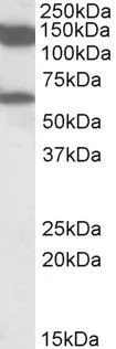
Figure 1. Western blot analysis of MLLT10 using anti-MLLT10 antibody (A04471-2). Electrophoresis was performed on a 5-20% SDS-PAGE gel at 70V (Stacking gel) / 90V (Resolving gel) for 2-3 hours. The sample well of each lane was loaded with 30 ug of sample under reducing conditions. Lane 1: rat testis tissue lysates, Lane 2: mouse testis tissue lysates. After electrophoresis, proteins were transferred to a nitrocellulose membrane at 150 mA for 50-90 minutes. Blocked the membrane with 5% non-fat milk/TBS for 1.5 hour at RT. The membrane was incubated with rabbit anti-MLLT10 antigen affinity purified polyclonal antibody (Catalog # A04471-2) at 0.5 microg/mL overnight at 4°C, then washed with TBS-0.1%Tween 3 times with 5 minutes each and probed with a goat anti-rabbit IgG-HRP secondary antibody at a dilution of 1:5000 for 1.5 hour at RT. The signal is developed using an Enhanced Chemiluminescent detection (ECL) kit (Catalog # EK1002) with Tanon 5200 system. A specific band was detected for MLLT10 at approximately 130 kDa. The expected band size for MLLT10 is at 113 kDa.
 and anti-Beta Tubulin antibody (M01857-3). MLLT10 was detected in immunocytochemical section of A549 cell. Enzyme antigen retrieval was performed using IHC enzyme antigen retrieval reagent (AR0022) for 15 mins. The cells were blocked with 10% goat serum. And then incubated with 5 microg/mL rabbit anti-MLLT10 Antibody (A04471-2) and mouse anti-Beta Tubulin antibody (M01857-3) overnight at 4°C. Cy3 Conjugated Goat Anti-Rabbit IgG (BA1032) and DyLight®488 Conjugated Goat Anti-Mouse IgG (BA1126) were used as secondary antibody at 1:500 dilution and incubated for 30 minutes at 37°C. Visualize using a fluorescence microscope and filter sets appropriate for the label used.")
. Overlay histogram showing PC-3 cells stained with A04471-2 (Blue line). To facilitate intracellular staining, cells were fixed with 4% paraformaldehyde and permeabilized with permeabilization buffer. The cells were blocked with 10% normal goat serum. And then incubated with rabbit anti-MLLT10 Antibody (A04471-2, 1 microg/1x106 cells) for 30 min at 20°C. DyLight®488 conjugated goat anti-rabbit IgG (BA1127, 5-10 microg/1x106 cells) was used as secondary antibody for 30 minutes at 20°C. Isotype control antibody (Green line) was rabbit IgG (1 microg/1x106) used under the same conditions. Unlabelled sample (Red line) was also used as a control.")
Figure 1. Western blot analysis of MLLT10 using anti-MLLT10 antibody (A04471-2). Electrophoresis was performed on a 5-20% SDS-PAGE gel at 70V (Stacking gel) / 90V (Resolving gel) for 2-3 hours. The sample well of each lane was loaded with 30 ug of sample under reducing conditions. Lane 1: rat testis tissue lysates, Lane 2: mouse testis tissue lysates. After electrophoresis, proteins were transferred to a nitrocellulose membrane at 150 mA for 50-90 minutes. Blocked the membrane with 5% non-fat milk/TBS for 1.5 hour at RT. The membrane was incubated with rabbit anti-MLLT10 antigen affinity purified polyclonal antibody (Catalog # A04471-2) at 0.5 microg/mL overnight at 4°C, then washed with TBS-0.1%Tween 3 times with 5 minutes each and probed with a goat anti-rabbit IgG-HRP secondary antibody at a dilution of 1:5000 for 1.5 hour at RT. The signal is developed using an Enhanced Chemiluminescent detection (ECL) kit (Catalog # EK1002) with Tanon 5200 system. A specific band was detected for MLLT10 at approximately 130 kDa. The expected band size for MLLT10 is at 113 kDa.
Anti-MLLT10 Antibody Picoband(r)
A04471-2-CARRIER-FREE
ApplicationsFlow Cytometry, ImmunoFluorescence, Western Blot, ELISA, ImmunoCytoChemistry
Product group Antibodies
ReactivityHuman, Mouse, Rat
TargetMLLT10
Overview
- SupplierBoster Bio
- Product NameAnti-MLLT10 Antibody Picoband(r)
- Delivery Days Customer9
- ApplicationsFlow Cytometry, ImmunoFluorescence, Western Blot, ELISA, ImmunoCytoChemistry
- CertificationResearch Use Only
- ClonalityPolyclonal
- Concentration500 ug/ml
- Gene ID8028
- Target nameMLLT10
- Target descriptionMLLT10 histone lysine methyltransferase DOT1L cofactor
- Target synonymsAF10, protein AF-10, ALL1-fused gene from chromosome 10 protein, myeloid/lymphoid or mixed-lineage leukemia (trithorax homolog, Drosophila); translocated to, 10, myeloid/lymphoid or mixed-lineage leukemia; translocated to, 10, type I AF10 protein, type III AF10 protein, type IV AF10 protein
- HostRabbit
- IsotypeIgG
- Protein IDP55197
- Protein NameProtein AF-10
- Scientific DescriptionBoster Bio Anti-MLLT10 Antibody Picoband® catalog # A04471-2. Tested in ELISA, IF, ICC, WB, Flow Cytometry applications. This antibody reacts with Human, Mouse, Rat. The brand Picoband indicates this is a premium antibody that guarantees superior quality, high affinity, and strong signals with minimal background in Western blot applications. Only our best-performing antibodies are designated as Picoband, ensuring unmatched performance.
- ReactivityHuman, Mouse, Rat
- Storage Instruction-20°C,2°C to 8°C
- UNSPSC12352203
Related products

Product group Antibodies

MLLT10 AntibodyLS-C749264
ApplicationsWestern Blot
ReactivityHuman, Mouse
TargetMLLT10
- SizePrice
Product group Antibodies
AF10 Polyclonal AntibodyBS-3696R
ApplicationsFlow Cytometry, Western Blot, ImmunoHistoChemistry, ImmunoHistoChemistry Frozen, ImmunoHistoChemistry Paraffin
ReactivityBovine, Canine, Equine, Human, Mouse, Porcine, Rabbit, Rat, Sheep
TargetMLLT10
- SizePrice
Product group Antibodies
MLLT10 AntibodyCSB-PA000830
ApplicationsImmunoFluorescence, Western Blot, ELISA, ImmunoHistoChemistry
ReactivityHuman, Mouse
TargetMLLT10
- SizePrice
Product group Antibodies

Goat anti-MLLT10 / AF10EB09975
ApplicationsWestern Blot, ELISA
ReactivityBovine, Canine, Human, Mouse, Rat
TargetMLLT10
- SizePrice
Product group Antibodies
AF10 antibody, InternalGTX88120
ApplicationsWestern Blot
ReactivityHuman, Rat
TargetMLLT10
- SizePrice

Product group Antibodies
Anti-MLLT10 AntibodyHPA005747
ApplicationsChIP Chromatin ImmunoPrecipitation, ImmunoCytoChemistry, ImmunoHistoChemistry
ReactivityHuman
TargetMLLT10
- SizePrice