


Immunohistochemical staining of human esophagus shows nuclear and cytoplasmic positivity in squamous epithelial cells.

Immunohistochemical staining of human esophagus shows nuclear and cytoplasmic positivity in squamous epithelial cells.
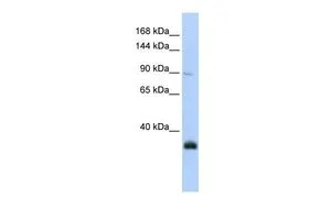
Anti-MORC3 Antibody
HPA018406
ApplicationsWestern Blot, ImmunoHistoChemistry
Product group Antibodies
ReactivityHuman
TargetMORC3
Overview
- SupplierAtlas Antibodies
- Product NameAnti-MORC3 Antibody
- Delivery Days Customer4
- ApplicationsWestern Blot, ImmunoHistoChemistry
- CertificationResearch Use Only
- ClonalityPolyclonal
- ConjugateUnconjugated
- Gene ID23515
- Target nameMORC3
- Target descriptionMORC family CW-type zinc finger 3
- Target synonymsNXP2, ZCW5, ZCWCC3, MORC family CW-type zinc finger protein 3, nuclear matrix protein 2, nuclear matrix protein NXP2, zinc finger CW-type coiled-coil domain protein 3, zinc finger, CW type with coiled-coil domain 3
- HostRabbit
- IsotypeIgG
- Protein IDQ14149
- Protein NameMORC family CW-type zinc finger protein 3
- Scientific DescriptionRecombinant Protein Epitope Signature Tag (PrEST) antigen sequence
- ReactivityHuman
- Storage Instruction-20°C,2°C to 8°C
- UNSPSC41116161
Datasheet
MSDS
Related products
Product group Antibodies

NXP2 / MORC3 AntibodyLS-C750386
ApplicationsWestern Blot
ReactivityHuman, Mouse
TargetMORC3
- SizePrice
Product group Antibodies
MORC3 antibody, N-termGTX46520
ApplicationsWestern Blot
ReactivityHuman
TargetMORC3
- SizePrice

Product group Antibodies
Anti-MORC3 AntibodyHPA034848
ApplicationsImmunoCytoChemistry
ReactivityHuman
TargetMORC3
- SizePrice
Product group Antibodies
MORC3 AntibodyCSB-PA169349XA01DOA
ApplicationsWestern Blot, ELISA
ReactivityPlant
- SizePrice

Product group Antibodies
Anti-MORC3 Antibody Picoband(r)A06075-4-CARRIER-FREE
ApplicationsFlow Cytometry, ImmunoFluorescence, Western Blot, ImmunoCytoChemistry, ImmunoHistoChemistry
ReactivityHuman, Mouse, Rat
TargetMORC3
- SizePrice