


Immunohistochemical staining of human testis shows moderate cytoplasmic positivity in cells in seminiferous ducts.
Immunohistochemical staining of human testis shows moderate cytoplasmic positivity in cells in seminiferous ducts.
Anti-MPHOSPH9 Antibody
HPA037485
ApplicationsImmunoHistoChemistry
Product group Antibodies
ReactivityHuman
TargetMPHOSPH9
Overview
- SupplierAtlas Antibodies
- Product NameAnti-MPHOSPH9 Antibody
- Delivery Days Customer4
- ApplicationsImmunoHistoChemistry
- CertificationResearch Use Only
- ClonalityPolyclonal
- ConjugateUnconjugated
- Gene ID10198
- Target nameMPHOSPH9
- Target descriptionM-phase phosphoprotein 9
- Target synonymsMPP-9, MPP9, M-phase phosphoprotein 9
- HostRabbit
- IsotypeIgG
- Protein IDQ99550
- Protein NameM-phase phosphoprotein 9
- Scientific DescriptionRecombinant Protein Epitope Signature Tag (PrEST) antigen sequence
- ReactivityHuman
- Storage Instruction-20°C,2°C to 8°C
- UNSPSC41116161
Datasheet
MSDS
Related products
Product group Antibodies
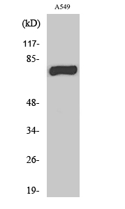
Anti-MPHOSPH9 AntibodyA99960
ApplicationsWestern Blot, ELISA
ReactivityHuman
- SizePrice
Product group Antibodies
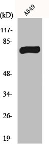
Anti-MPHOSPH9 (N-term) Antibody102-23041
ApplicationsWestern Blot
TargetMPHOSPH9
- SizePrice

Product group Antibodies
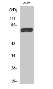
Anti-MPHOSPH9 Antibody Picoband(r)A11350-2-CARRIER-FREE
ApplicationsFlow Cytometry, ImmunoFluorescence, Western Blot, ELISA, ImmunoCytoChemistry
ReactivityHuman, Mouse, Rat
TargetMPHOSPH9
- SizePrice
Product group Antibodies
MPHOSPH9 Polyclonal AntibodyBS-7812R
ApplicationsImmunoFluorescence, Western Blot, ELISA, ImmunoCytoChemistry, ImmunoHistoChemistry, ImmunoHistoChemistry Frozen, ImmunoHistoChemistry Paraffin
ReactivityCanine, Human, Mouse, Rat
TargetMPHOSPH9
- SizePrice
Product group Antibodies
MPHOSPH9 AntibodyCSB-PA003268
ApplicationsWestern Blot, ELISA
ReactivityHuman
TargetMPHOSPH9
- SizePrice
Product group Antibodies

ApplicationsWestern Blot, ELISA
ReactivityHuman
TargetMPHOSPH9
- SizePrice
Product group Antibodies
MPHOSPH9 antibodyGTX34083
ApplicationsWestern Blot
ReactivityHuman
TargetMPHOSPH9
- SizePrice
Product group Antibodies
Anti-MPHOSPH9Y158492
ApplicationsWestern Blot, ELISA, ImmunoHistoChemistry
ReactivityHuman
- SizePrice