

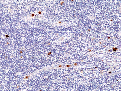
Immunohistochemical staining of formalin fixed and paraffin embedded human tonsil tissue section using Anti-CD23 Rabbit Monoclonal Antibody (Clone RM406) at a 1:200 dilution.
Immunohistochemical staining of formalin fixed and paraffin embedded human tonsil tissue section using Anti-CD23 Rabbit Monoclonal Antibody (Clone RM406) at a 1:200 dilution.
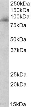
anti-MPO (human), Rabbit Monoclonal (RM407)
REV-31-1293-00
ApplicationsWestern Blot, ImmunoHistoChemistry
Product group Antibodies
ReactivityHuman
TargetMPO
Overview
- SupplierRevMAb Biosciences
- Product Nameanti-MPO (human), Rabbit Monoclonal (RM407)
- Delivery Days Customer10
- ApplicationsWestern Blot, ImmunoHistoChemistry
- CertificationResearch Use Only
- ClonalityMonoclonal
- Clone IDRM407
- Gene ID4353
- Target nameMPO
- Target descriptionmyeloperoxidase
- Target synonymsmyeloperoxidase
- HostRabbit
- IsotypeIgG
- Protein IDP05164
- Protein NameMyeloperoxidase
- Scientific DescriptionMyeloperoxidase (MPO) is a hemoprotein that is abundantly expressed in neutrophils and secreted during their activation. Native Myeloperoxidase is represented as a covalently bound tetrameric complex of two glycosylated alpha chains and two unglycosylated beta chains. Traditionally, myeloperoxidase was considered as a main target of anti-neutrophil cytoplasm antibodies (ANCA), the serological markers for certain systemic vasculities such as periarteriitis nodosa, microscopic polyarteriitis and pulmonary eosinophilic granulomatosis (Churg-Strauss syndrome). Low to moderate anti-myeloperoxidase autoantibody levels are also reported in rheumatoid arthritis. Myeloperoxidase also participates in the initiation and progression of cardiovascular disease and is a promising cardiac markers. Myeloperoxidase possesses potent proinflammatory properties and may contribute directly to tissue injury. Myeloperoxidase is part of the host defense system of polymorphonuclear leukocytes and myeloperoxidase is responsible for microbicidal activity against a wide range of organisms. In stimulated PMN (polymorphonuclear leukocytes), MPO catalyzes the production of hypohalous acids, primarily hypochlorous acid in physiologic situations, and other toxic intermediates that greatly enhance PMN microbicidal activity. Myeloperoxidase immunostaining is important in the diagnosis of myeloid sarcoma, contrasting with the negative staining of lymphomas. - Recombinant Antibody. This antibody reacts to human to MPO. Applications: WB, IHC. Source: Rabbit. Liquid. 50% Glycerol/PBS with 1% BSA and 0.09% sodium azide. Myeloperoxidase (MPO) is a hemoprotein that is abundantly expressed in neutrophils and secreted during their activation. Native Myeloperoxidase is represented as a covalently bound tetrameric complex of two glycosylated alpha chains and two unglycosylated beta chains. Traditionally, myeloperoxidase was considered as a main target of anti-neutrophil cytoplasm antibodies (ANCA), the serological markers for certain systemic vasculities such as periarteriitis nodosa, microscopic polyarteriitis and pulmonary eosinophilic granulomatosis (Churg-Strauss syndrome). Low to moderate anti-myeloperoxidase autoantibody levels are also reported in rheumatoid arthritis. Myeloperoxidase also participates in the initiation and progression of cardiovascular disease and is a promising cardiac markers. Myeloperoxidase possesses potent proinflammatory properties and may contribute directly to tissue injury. Myeloperoxidase is part of the host defense system of polymorphonuclear leukocytes and myeloperoxidase is responsible for microbicidal activity against a wide range of organisms. In stimulated PMN (polymorphonuclear leukocytes), MPO catalyzes the production of hypohalous acids, primarily hypochlorous acid in physiologic situations, and other toxic intermediates that greatly enhance PMN microbicidal activity. Myeloperoxidase immunostaining is important in the diagnosis of myeloid sarcoma, contrasting with the negative staining of lymphomas.
- ReactivityHuman
- Storage Instruction-20°C,2°C to 8°C
- UNSPSC41116161
Datasheet
Related products
Product group Antibodies
ApplicationsWestern Blot, ELISA
ReactivityHuman
- SizePrice
Product group Antibodies

Anti-MPO [12F3A4C11]Ab03400-10.0
ApplicationsWestern Blot, ImmunoHistoChemistry
ReactivityHuman
TargetMPO
- SizePrice
Product group Antibodies
Anti-MPO Antibody144-62885
ApplicationsImmunoFluorescence, Western Blot, ImmunoHistoChemistry
ReactivityHuman, Mouse, Rat
TargetMPO
- SizePrice
Product group Antibodies
Anti-MPO AntibodyAMAB91997
ApplicationsWestern Blot, ImmunoHistoChemistry
ReactivityHuman
TargetMPO
- SizePrice

Product group Antibodies
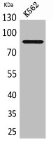
Anti-MPO Antibody Picoband(r)A00372-1-CARRIER-FREE
ApplicationsFlow Cytometry, Western Blot, ELISA
ReactivityHuman
TargetMPO
- SizePrice
Product group Antibodies
References
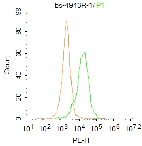
MPO Polyclonal AntibodyBS-4943R
ApplicationsFlow Cytometry, ImmunoFluorescence, ELISA, ImmunoCytoChemistry, ImmunoHistoChemistry, ImmunoHistoChemistry Frozen, ImmunoHistoChemistry Paraffin
ReactivityCanine, Equine, Guinea Pig, Human, Mouse, Rabbit, Rat
TargetMPO
- SizePrice
Product group Antibodies

Goat anti-myeloperoxidaseEB11953
ApplicationsWestern Blot, ELISA
ReactivityHuman
TargetMPO
- SizePrice
Product group Antibodies
Mpo Polyclonal AntibodyCAC09088
ApplicationsImmunoFluorescence, ELISA, ImmunoHistoChemistry
TargetMPO
- SizePrice
Product group Antibodies
MPO AntibodyCSB-PA005559
ApplicationsWestern Blot, ELISA
ReactivityHuman, Mouse, Rat
TargetMPO
- SizePrice