


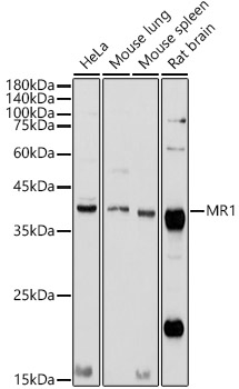
Figure 1. Western blot analysis of MR1 using anti-MR1 antibody (A00618-1). Electrophoresis was performed on a 5-20% SDS-PAGE gel at 70V (Stacking gel) / 90V (Resolving gel) for 2-3 hours. The sample well of each lane was loaded with 50ug of sample under reducing conditions. Lane 1: human T-47D whole cell lysates, Lane 2: human U-937 whole cell lysates, Lane 3: human A431 whole cell lysates. After Electrophoresis, proteins were transferred to a Nitrocellulose membrane at 150mA for 50-90 minutes. Blocked the membrane with 5% Non-fat Milk/ TBS for 1.5 hour at RT. The membrane was incubated with rabbit anti-MR1 antigen affinity purified polyclonal antibody (Catalog # A00618-1) at 0.5 microg/mL overnight at 4°C, then washed with TBS-0.1%Tween 3 times with 5 minutes each and probed with a goat anti-rabbit IgG-HRP secondary antibody at a dilution of 1:10000 for 1.5 hour at RT. The signal is developed using an Enhanced Chemiluminescent detection (ECL) kit (Catalog # EK1002) with Tanon 5200 system. A specific band was detected for MR1 at approximately 40KD. The expected band size for MR1 is at 43KD.
. Overlay histogram showing SiHa cells stained with A00618-1 (Blue line).The cells were blocked with 10% normal goat serum. And then incubated with rabbit anti-MR1 Antibody (A00618-1,1microg/1x106 cells) for 30 min at 20°C. DyLight®488 conjugated goat anti-rabbit IgG (BA1127, 5-10microg/1x106 cells) was used as secondary antibody for 30 minutes at 20°C. Isotype control antibody (Green line) was rabbit IgG (1microg/1x106) used under the same conditions. Unlabelled sample (Red line) was also used as a control.")
Figure 1. Western blot analysis of MR1 using anti-MR1 antibody (A00618-1). Electrophoresis was performed on a 5-20% SDS-PAGE gel at 70V (Stacking gel) / 90V (Resolving gel) for 2-3 hours. The sample well of each lane was loaded with 50ug of sample under reducing conditions. Lane 1: human T-47D whole cell lysates, Lane 2: human U-937 whole cell lysates, Lane 3: human A431 whole cell lysates. After Electrophoresis, proteins were transferred to a Nitrocellulose membrane at 150mA for 50-90 minutes. Blocked the membrane with 5% Non-fat Milk/ TBS for 1.5 hour at RT. The membrane was incubated with rabbit anti-MR1 antigen affinity purified polyclonal antibody (Catalog # A00618-1) at 0.5 microg/mL overnight at 4°C, then washed with TBS-0.1%Tween 3 times with 5 minutes each and probed with a goat anti-rabbit IgG-HRP secondary antibody at a dilution of 1:10000 for 1.5 hour at RT. The signal is developed using an Enhanced Chemiluminescent detection (ECL) kit (Catalog # EK1002) with Tanon 5200 system. A specific band was detected for MR1 at approximately 40KD. The expected band size for MR1 is at 43KD.
Anti-MR1 Antibody Picoband(r)
A00618-1-CARRIER-FREE
ApplicationsFlow Cytometry, Western Blot, ELISA
Product group Antibodies
ReactivityHuman, Mouse, Rat
TargetMR1
Overview
- SupplierBoster Bio
- Product NameAnti-MR1 Antibody Picoband(r)
- Delivery Days Customer9
- ApplicationsFlow Cytometry, Western Blot, ELISA
- CertificationResearch Use Only
- ClonalityPolyclonal
- Concentration500 ug/ml
- Gene ID3140
- Target nameMR1
- Target descriptionmajor histocompatibility complex, class I-related
- Target synonymsHLALS, major histocompatibility complex class I-related protein 1, MHC class I-like antigen MR-1, MHC class I-related protein, MHC class I-related protein 1, MHC class-I related-gene protein, major histocompatibility complex class I-related gene protein, major histocompatibility complex, class I-like sequence
- HostRabbit
- IsotypeIgG
- Protein IDQ95460
- Protein NameMajor histocompatibility complex class I-related protein 1
- Scientific DescriptionBoster Bio Anti-MR1 Antibody catalog # A00618-1. Tested in ELISA, Flow Cytometry, WB applications. This antibody reacts with Human, Mouse, Rat. The brand Picoband indicates this is a premium antibody that guarantees superior quality, high affinity, and strong signals with minimal background in Western blot applications. Only our best-performing antibodies are designated as Picoband, ensuring unmatched performance.
- ReactivityHuman, Mouse, Rat
- Storage Instruction-20°C,2°C to 8°C
- UNSPSC12352203
Related products
Product group Antibodies
Anti-MR1 AntibodyA10277
ApplicationsImmunoFluorescence, Western Blot, ImmunoCytoChemistry
ReactivityHuman, Mouse, Rat
- SizePrice
Product group Antibodies
Anti-MR1 Antibody144-66535
ApplicationsWestern Blot
ReactivityHuman, Mouse
TargetMR1
- SizePrice
Product group Antibodies

MR1 AntibodyLS-C830485
ApplicationsELISA, ImmunoHistoChemistry
ReactivityHuman
TargetMR1
- SizePrice
Product group Antibodies
MR1 AntibodyCSB-PA853268ESR2HU
ApplicationsELISA, ImmunoHistoChemistry
ReactivityHuman
TargetMR1
- SizePrice
Product group Antibodies
Mr1 Polyclonal AntibodyCAC07671
ApplicationsImmunoFluorescence, Western Blot, ELISA, ImmunoHistoChemistry
ReactivityMouse
TargetMR1
- SizePrice

Product group Antibodies
Anti-MR1 AntibodyHPA048304
ApplicationsImmunoCytoChemistry
ReactivityHuman
TargetMR1
- SizePrice
Product group Antibodies
Anti-MR1 AntibodyCAB8051
ApplicationsImmunoFluorescence, Western Blot, ELISA, ImmunoCytoChemistry
ReactivityHuman
TargetMR1
- SizePrice