

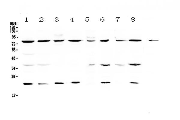
Figure 1. Western blot analysis of MRE11 using anti-MRE11 antibody (A00731-2). Electrophoresis was performed on a 5-20% SDS-PAGE gel at 70V (Stacking gel) / 90V (Resolving gel) for 2-3 hours. The sample well of each lane was loaded with 50ug of sample under reducing conditions. Lane 1: human Hela whole cell lysates, Lane 2: human MCF-7 whole cell lysates, Lane 3: human COLO-320 whole cell lysates, Lane 4: human U-87MG whole cell lysates, Lane 5: rat brain tissue lysates, Lane 6: rat liver tissue lysates, Lane 7: mouse brain tissue lysates, Lane 8: mouse liver tissue lysates. After Electrophoresis, proteins were transferred to a Nitrocellulose membrane at 150mA for 50-90 minutes. Blocked the membrane with 5% Non-fat Milk/ TBS for 1.5 hour at RT. The membrane was incubated with rabbit anti-MRE11 antigen affinity purified polyclonal antibody (Catalog # A00731-2) at 0.5 microg/mL overnight at 4°C, then washed with TBS-0.1%Tween 3 times with 5 minutes each and probed with a goat anti-rabbit IgG-HRP secondary antibody at a dilution of 1:10000 for 1.5 hour at RT. The signal is developed using an Enhanced Chemiluminescent detection (ECL) kit (Catalog # EK1002) with Tanon 5200 system. A specific band was detected for MRE11 at approximately 81KD. The expected band size for MRE11 is at 81KD.
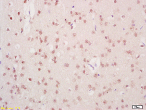
. MRE11 was detected in paraffin-embedded section of human colon cancer tissue. Heat mediated antigen retrieval was performed in citrate buffer (pH6, epitope retrieval solution) for 20 mins. The tissue section was blocked with 10% goat serum. The tissue section was then incubated with 1microg/ml rabbit anti-MRE11 Antibody (A00731-2) overnight at 4°C. Biotinylated goat anti-rabbit IgG was used as secondary antibody and incubated for 30 minutes at 37°C. The tissue section was developed using Strepavidin-Biotin-Complex (SABC)(Catalog # SA1022) with DAB as the chromogen.")
. MRE11 was detected in paraffin-embedded section of human lung cancer tissue. Heat mediated antigen retrieval was performed in citrate buffer (pH6, epitope retrieval solution) for 20 mins. The tissue section was blocked with 10% goat serum. The tissue section was then incubated with 1microg/ml rabbit anti-MRE11 Antibody (A00731-2) overnight at 4°C. Biotinylated goat anti-rabbit IgG was used as secondary antibody and incubated for 30 minutes at 37°C. The tissue section was developed using Strepavidin-Biotin-Complex (SABC)(Catalog # SA1022) with DAB as the chromogen.")
. MRE11 was detected in paraffin-embedded section of human mammary cancer tissue. Heat mediated antigen retrieval was performed in citrate buffer (pH6, epitope retrieval solution) for 20 mins. The tissue section was blocked with 10% goat serum. The tissue section was then incubated with 1microg/ml rabbit anti-MRE11 Antibody (A00731-2) overnight at 4°C. Biotinylated goat anti-rabbit IgG was used as secondary antibody and incubated for 30 minutes at 37°C. The tissue section was developed using Strepavidin-Biotin-Complex (SABC)(Catalog # SA1022) with DAB as the chromogen.")
Figure 1. Western blot analysis of MRE11 using anti-MRE11 antibody (A00731-2). Electrophoresis was performed on a 5-20% SDS-PAGE gel at 70V (Stacking gel) / 90V (Resolving gel) for 2-3 hours. The sample well of each lane was loaded with 50ug of sample under reducing conditions. Lane 1: human Hela whole cell lysates, Lane 2: human MCF-7 whole cell lysates, Lane 3: human COLO-320 whole cell lysates, Lane 4: human U-87MG whole cell lysates, Lane 5: rat brain tissue lysates, Lane 6: rat liver tissue lysates, Lane 7: mouse brain tissue lysates, Lane 8: mouse liver tissue lysates. After Electrophoresis, proteins were transferred to a Nitrocellulose membrane at 150mA for 50-90 minutes. Blocked the membrane with 5% Non-fat Milk/ TBS for 1.5 hour at RT. The membrane was incubated with rabbit anti-MRE11 antigen affinity purified polyclonal antibody (Catalog # A00731-2) at 0.5 microg/mL overnight at 4°C, then washed with TBS-0.1%Tween 3 times with 5 minutes each and probed with a goat anti-rabbit IgG-HRP secondary antibody at a dilution of 1:10000 for 1.5 hour at RT. The signal is developed using an Enhanced Chemiluminescent detection (ECL) kit (Catalog # EK1002) with Tanon 5200 system. A specific band was detected for MRE11 at approximately 81KD. The expected band size for MRE11 is at 81KD.
Anti-MRE11/MRE11 Antibody Picoband(r)
A00731-2-CARRIER-FREE
ApplicationsWestern Blot, ELISA, ImmunoHistoChemistry
Product group Antibodies
ReactivityHuman, Mouse, Rat
TargetMRE11
Overview
- SupplierBoster Bio
- Product NameAnti-MRE11/MRE11 Antibody Picoband(r)
- Delivery Days Customer9
- ApplicationsWestern Blot, ELISA, ImmunoHistoChemistry
- CertificationResearch Use Only
- ClonalityPolyclonal
- Concentration500 ug/ml
- Gene ID4361
- Target nameMRE11
- Target descriptionMRE11 double strand break repair nuclease
- Target synonymsATLD, HNGS1, MRE11A, MRE11B, double-strand break repair protein MRE11, AT-like disease, DNA recombination and repair protein, MRE11 double strand break repair nuclease A, MRE11 homolog 1, MRE11 homolog A, double strand break repair nuclease, MRE11 homolog, double strand break repair nuclease A, MRE11 meiotic recombination 11 homolog A, MRE11 meiotic recombination 11-like protein A, double-strand break repair protein MRE11A, endo/exonuclease Mre11, meiotic recombination 11 homolog 1, meiotic recombination 11 homolog A
- HostRabbit
- IsotypeIgG
- Protein IDP49959
- Protein NameDouble-strand break repair protein MRE11
- Scientific DescriptionBoster Bio Anti-MRE11/MRE11 Antibody Picoband® catalog # A00731-2. Tested in ELISA, IHC, WB applications. This antibody reacts with Human, Mouse, Rat. The brand Picoband indicates this is a premium antibody that guarantees superior quality, high affinity, and strong signals with minimal background in Western blot applications. Only our best-performing antibodies are designated as Picoband, ensuring unmatched performance.
- ReactivityHuman, Mouse, Rat
- Storage Instruction-20°C,2°C to 8°C
- UNSPSC12352203
Related products
Product group Antibodies
MRE11A AntibodyCSB-PA003273
ApplicationsWestern Blot, ELISA, ImmunoHistoChemistry
ReactivityHuman, Monkey, Mouse, Rat
TargetMRE11
- SizePrice
Product group Antibodies
ApplicationsImmunoPrecipitation, Western Blot, ImmunoCytoChemistry, ImmunoHistoChemistry
ReactivityMouse, Rat
TargetMRE11
- SizePrice
Product group Antibodies
Anti-MRE11A Antibody144-02559
ApplicationsImmunoFluorescence, ImmunoPrecipitation, Western Blot
ReactivityHuman, Mouse, Rat
TargetMRE11
- SizePrice
Product group Antibodies
Anti-MRE11A AntibodyA96825
ApplicationsWestern Blot, ELISA
ReactivityHuman, Mouse, Rat
- SizePrice

Product group Antibodies
Anti-MRE11 AntibodyHPA002691
ApplicationsWestern Blot, ImmunoCytoChemistry, ImmunoHistoChemistry
ReactivityHuman
TargetMRE11
- SizePrice
Product group Antibodies

MRE11A / MRE11 AntibodyLS-C401850
ApplicationsWestern Blot, ELISA, ImmunoHistoChemistry
ReactivityHuman, Mouse, Rat
TargetMRE11
- SizePrice
Product group Antibodies
Mre11 Polyclonal AntibodyBS-3503R
ApplicationsImmunoFluorescence, Western Blot, ELISA, ImmunoCytoChemistry, ImmunoHistoChemistry, ImmunoHistoChemistry Frozen, ImmunoHistoChemistry Paraffin
ReactivityBovine, Canine, Equine, Human, Mouse, Rabbit, Rat
TargetMRE11
- SizePrice
![Non-transfected (–) and transfected (+) HeLa whole cell extracts (30 μg) were separated by 7.5% SDS-PAGE, and the membrane was blotted with Mre11 antibody [C1C3-2] (GTX111814) diluted at 1:1000.](https://www.genetex.com/upload/website/prouct_img/normal/GTX111814/GTX111814_40492_20160922_WB_shRNA_watermark_w_23060500_893.webp)
Product group Antibodies
Mre11 antibody [C1C3-2]GTX111814
ApplicationsImmunoFluorescence, ImmunoPrecipitation, Western Blot, ImmunoCytoChemistry
ReactivityHuman
TargetMRE11
- SizePrice