

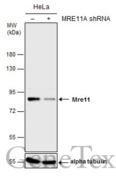
Non-transfected (–) and transfected (+) HeLa whole cell extracts (30 μg) were separated by 7.5% SDS-PAGE, and the membrane was blotted with Mre11 antibody [C1C3-2] (GTX111814) diluted at 1:1000.
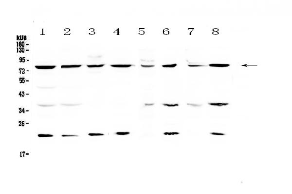
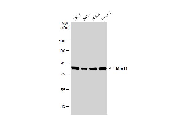
![Various whole cell extracts (30 μg) were separated by 7.5% SDS-PAGE, and the membranes were blotted with Mre11 antibody [C1C3-2] (GTX111814) diluted at 1:1000 and competitor's antibody diluted at 1:1000. The HRP-conjugated anti-rabbit IgG antibody (GTX213110-01) was used to detect the primary antibody. *The competitor is not affiliated with GeneTex and does not endorse this product.](https://www.genetex.com/upload/website/prouct_img/normal/GTX111814/GTX111814_40492_20200403_WB_competitor_watermark_w_23060500_716.webp "Various whole cell extracts (30 μg) were separated by 7.5% SDS-PAGE, and the membranes were blotted with Mre11 antibody [C1C3-2] (GTX111814) diluted at 1:1000 and competitor's antibody diluted at 1:1000. The HRP-conjugated anti-rabbit IgG antibody (GTX213110-01) was used to detect the primary antibody. *The competitor is not affiliated with GeneTex and does not endorse this product.")
![Mre11 antibody [C1C3-2] detects Mre11 protein at nucleus by immunofluorescent analysis. Sample: SKNSH cells were fixed in 4% paraformaldehyde at RT for 15 min. Green: Mre11 protein stained by Mre11 antibody [C1C3-2] (GTX111814) diluted at 1:500. Blue: Hoechst 33342 staining. Scale bar = 10 μm.](https://www.genetex.com/upload/website/prouct_img/normal/GTX111814/GTX111814_40492_IFA_w_23060500_978.webp "Mre11 antibody [C1C3-2] detects Mre11 protein at nucleus by immunofluorescent analysis. Sample: SKNSH cells were fixed in 4% paraformaldehyde at RT for 15 min. Green: Mre11 protein stained by Mre11 antibody [C1C3-2] (GTX111814) diluted at 1:500. Blue: Hoechst 33342 staining. Scale bar = 10 μm.")
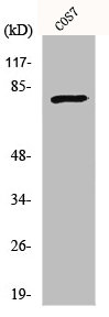
![Non-transfected (–) and transfected (+) 293T whole cell extracts (30 μg) were separated by 7.5% SDS-PAGE, and the membrane was blotted with Mre11 antibody [C1C3-2] (GTX111814) diluted at 1:1000. The HRP-conjugated anti-rabbit IgG antibody (GTX213110-01) was used to detect the primary antibody.](https://www.genetex.com/upload/website/prouct_img/normal/GTX111814/GTX111814_40492_20181026_WB_B_w_23060500_547.webp "Non-transfected (–) and transfected (+) 293T whole cell extracts (30 μg) were separated by 7.5% SDS-PAGE, and the membrane was blotted with Mre11 antibody [C1C3-2] (GTX111814) diluted at 1:1000. The HRP-conjugated anti-rabbit IgG antibody (GTX213110-01) was used to detect the primary antibody.")
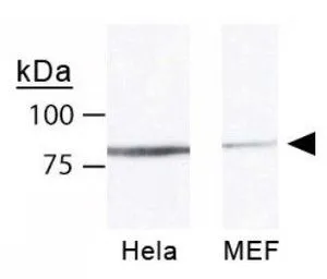
![Immunoprecipitation of Mre11 protein from HeLa whole cell extracts using 5 μg of Mre11 antibody [C1C3-2] (GTX111814). Western blot analysis was performed using Mre11 antibody [C1C3-2] (GTX111814). EasyBlot anti-Rabbit IgG (GTX221666-01) was used as a secondary reagent.](https://www.genetex.com/upload/website/prouct_img/normal/GTX111814/GTX111814_40492_20151106_IP_w_23060500_551.webp "Immunoprecipitation of Mre11 protein from HeLa whole cell extracts using 5 μg of Mre11 antibody [C1C3-2] (GTX111814). Western blot analysis was performed using Mre11 antibody [C1C3-2] (GTX111814). EasyBlot anti-Rabbit IgG (GTX221666-01) was used as a secondary reagent.")
Non-transfected (–) and transfected (+) HeLa whole cell extracts (30 μg) were separated by 7.5% SDS-PAGE, and the membrane was blotted with Mre11 antibody [C1C3-2] (GTX111814) diluted at 1:1000.
Mre11 antibody [C1C3-2]
GTX111814
ApplicationsImmunoFluorescence, ImmunoPrecipitation, Western Blot, ImmunoCytoChemistry
Product group Antibodies
ReactivityHuman
TargetMRE11
Overview
- SupplierGeneTex
- Product NameMre11 antibody [C1C3-2]
- Delivery Days Customer9
- Application Supplier NoteWB: 1:500-1:3000. ICC/IF: 1:100-1:1000. IP: 1:100-1:500. *Optimal dilutions/concentrations should be determined by the researcher.Not tested in other applications.
- ApplicationsImmunoFluorescence, ImmunoPrecipitation, Western Blot, ImmunoCytoChemistry
- CertificationResearch Use Only
- ClonalityPolyclonal
- Concentration0.57 mg/ml
- ConjugateUnconjugated
- Gene ID4361
- Target nameMRE11
- Target descriptionMRE11 double strand break repair nuclease
- Target synonymsATLD, HNGS1, MRE11A, MRE11B, double-strand break repair protein MRE11, AT-like disease, DNA recombination and repair protein, MRE11 double strand break repair nuclease A, MRE11 homolog 1, MRE11 homolog A, double strand break repair nuclease, MRE11 homolog, double strand break repair nuclease A, MRE11 meiotic recombination 11 homolog A, MRE11 meiotic recombination 11-like protein A, double-strand break repair protein MRE11A, endo/exonuclease Mre11, meiotic recombination 11 homolog 1, meiotic recombination 11 homolog A
- HostRabbit
- IsotypeIgG
- Protein IDP49959
- Protein NameDouble-strand break repair protein MRE11
- Scientific DescriptionThis gene encodes a nuclear protein involved in homologous recombination, telomere length maintenance, and DNA double-strand break repair. By itself, the protein has 3 to 5 exonuclease activity and endonuclease activity. The protein forms a complex with the RAD50 homolog; this complex is required for nonhomologous joining of DNA ends and possesses increased single-stranded DNA endonuclease and 3 to 5 exonuclease activities. In conjunction with a DNA ligase, this protein promotes the joining of noncomplementary ends in vitro using short homologies near the ends of the DNA fragments. This gene has a pseudogene on chromosome 3. Alternative splicing of this gene results in two transcript variants encoding different isoforms. [provided by RefSeq]
- ReactivityHuman
- Storage Instruction-20°C or -80°C,2°C to 8°C
- UNSPSC41116161
Datasheet
Related products
Product group Antibodies
Anti-MRE11A AntibodyA96825
ApplicationsWestern Blot, ELISA
ReactivityHuman, Mouse, Rat
- SizePrice
Product group Antibodies
Anti-MRE11/MRE11 Antibody Picoband(r)A00731-2-CARRIER-FREE
ApplicationsWestern Blot, ELISA, ImmunoHistoChemistry
ReactivityHuman, Mouse, Rat
TargetMRE11
- SizePrice
Product group Antibodies
Anti-MRE11A Antibody144-02559
ApplicationsImmunoFluorescence, ImmunoPrecipitation, Western Blot
ReactivityHuman, Mouse, Rat
TargetMRE11
- SizePrice
Product group Antibodies
Mre11 Polyclonal AntibodyBS-3503R
ApplicationsImmunoFluorescence, Western Blot, ELISA, ImmunoCytoChemistry, ImmunoHistoChemistry, ImmunoHistoChemistry Frozen, ImmunoHistoChemistry Paraffin
ReactivityBovine, Canine, Equine, Human, Mouse, Rabbit, Rat
TargetMRE11
- SizePrice
Product group Antibodies
MRE11A AntibodyCSB-PA003273
ApplicationsWestern Blot, ELISA, ImmunoHistoChemistry
ReactivityHuman, Monkey, Mouse, Rat
TargetMRE11
- SizePrice
Product group Antibodies
ApplicationsImmunoPrecipitation, Western Blot, ImmunoCytoChemistry, ImmunoHistoChemistry
ReactivityMouse, Rat
TargetMRE11
- SizePrice
Product group Antibodies

MRE11A / MRE11 AntibodyLS-C401850
ApplicationsWestern Blot, ELISA, ImmunoHistoChemistry
ReactivityHuman, Mouse, Rat
TargetMRE11
- SizePrice
Product group Antibodies
References
Mre11 antibodyGTX30294
ApplicationsFlow Cytometry, ImmunoFluorescence, ImmunoPrecipitation, Western Blot, ChIP Chromatin ImmunoPrecipitation, ImmunoCytoChemistry, ImmunoHistoChemistry, ImmunoHistoChemistry Frozen, ImmunoHistoChemistry Paraffin
ReactivityChicken, Hamster, Human, Mouse, Rat
TargetMRE11
- SizePrice

Product group Antibodies
Anti-MRE11 AntibodyHPA002691
ApplicationsWestern Blot, ImmunoCytoChemistry, ImmunoHistoChemistry
ReactivityHuman
TargetMRE11
- SizePrice
Product group Antibodies
Mre11 antibodyGTX118741
ApplicationsImmunoFluorescence, ImmunoPrecipitation, Western Blot, ImmunoCytoChemistry, ImmunoHistoChemistry, ImmunoHistoChemistry Paraffin
ReactivityHuman, Mouse, Rat
TargetMRE11
- SizePrice