


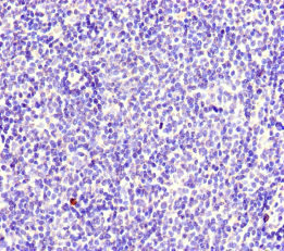
Immunohistochemistry analysis in human bone marrow and cerebral cortex tissues using Anti-MYO1G antibody. Corresponding MYO1G RNA-seq data are presented for the same tissues.

Immunohistochemistry analysis in human bone marrow and cerebral cortex tissues using Anti-MYO1G antibody. Corresponding MYO1G RNA-seq data are presented for the same tissues.
Anti-MYO1G Antibody
HPA021252
ApplicationsWestern Blot, ImmunoHistoChemistry
Product group Antibodies
ReactivityHuman
TargetMYO1G
Overview
- SupplierAtlas Antibodies
- Product NameAnti-MYO1G Antibody
- Delivery Days Customer4
- ApplicationsWestern Blot, ImmunoHistoChemistry
- CertificationResearch Use Only
- ClonalityPolyclonal
- ConjugateUnconjugated
- Gene ID64005
- Target nameMYO1G
- Target descriptionmyosin IG
- Target synonymsHA2, HLA-HA2, MHAG, unconventional myosin-Ig, minor histocompatibility antigen HA-2, myosin-Ig
- HostRabbit
- IsotypeIgG
- Protein IDB0I1T2
- Protein NameUnconventional myosin-Ig
- Scientific DescriptionRecombinant Protein Epitope Signature Tag (PrEST) antigen sequence
- ReactivityHuman
- Storage Instruction-20°C,2°C to 8°C
- UNSPSC41116161
Datasheet
MSDS
Related products
Product group Antibodies
MYO1G AntibodyPACO46046
ApplicationsImmunoFluorescence, ELISA, ImmunoHistoChemistry
ReactivityHuman
TargetMYO1G
- SizePrice
Product group Antibodies
ApplicationsImmunoPrecipitation, Western Blot, ImmunoCytoChemistry, ImmunoHistoChemistry
ReactivityRat
TargetMYO1G
- SizePrice

Product group Antibodies
Anti-MYO1G Antibody Picoband(r)A08448-2-CARRIER-FREE
ApplicationsFlow Cytometry, Western Blot, ELISA
ReactivityHuman
TargetMYO1G
- SizePrice
Product group Antibodies
MYO1G AntibodyCSB-PA015344LA01HU
ApplicationsImmunoFluorescence, ELISA, ImmunoHistoChemistry
ReactivityHuman
TargetMYO1G
- SizePrice
Product group Antibodies

MYO1G / HA2 Antibody (Biotin)LS-C678732
ApplicationsELISA
ReactivityHuman
TargetMYO1G
- SizePrice