

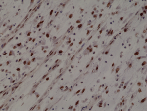
Immunohistochemical staining of formalin fixed and paraffin embedded human Rhabdomyosarcoma section using anti-MyoD1 rabbit monoclonal antibody (Clone RM369) at a 1:1000 dilution.
Immunohistochemical staining of formalin fixed and paraffin embedded human Rhabdomyosarcoma section using anti-MyoD1 rabbit monoclonal antibody (Clone RM369) at a 1:1000 dilution.
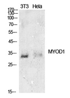
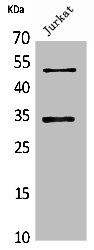
anti-MyoD1 (human), Rabbit Monoclonal (RM369)
REV-31-1255-00
ApplicationsWestern Blot, ImmunoHistoChemistry
Product group Antibodies
ReactivityHuman
TargetMYOD1
Overview
- SupplierRevMAb Biosciences
- Product Nameanti-MyoD1 (human), Rabbit Monoclonal (RM369)
- Delivery Days Customer2
- ApplicationsWestern Blot, ImmunoHistoChemistry
- CertificationResearch Use Only
- ClonalityMonoclonal
- Clone IDRM369
- Gene ID4654
- Target nameMYOD1
- Target descriptionmyogenic differentiation 1
- Target synonymsCMYO17, CMYP17, MYF3, MYOD, MYODRIF, PUM, bHLHc1, myoblast determination protein 1, class C basic helix-loop-helix protein 1, myf-3, myogenic factor 3
- HostRabbit
- IsotypeIgG
- Protein IDP15172
- Protein NameMyoblast determination protein 1
- Scientific DescriptionMyoD (MyoD1; Myoblast Determination Protein 1) plays a major role in regulating muscle differentiation. It belongs to a family of proteins known as myogenic regulatory factors (MRFs). These bHLH (basic helix loop helix) transcription factors act sequentially in myogenic differentiation. Vertebrate MRF family members include MyoD1, Myf5, myogenin and MRF4 (Myf6). MyoD is one of the earliest markers of myogenic commitment. MyoD is expressed at extremely low and essentially undetectable levels in quiescent satellite cells, but expression of MyoD is activated in response to exercise or muscle tissue damage. High MyoD expression represses cell renewal, promotes terminal differentiation and can induce apoptosis. The function of MyoD in development is to commit mesoderm cells to a skeletal myoblast lineage, and then to regulate that continued state. MyoD may also regulate muscle repair. MyoD mRNA levels are also reported to be elevated in aging skeletal muscle. One of the main actions of MyoD is to remove cells from the cell cycle (halt proliferation for terminal cell cycle arrest in differentiated myocytes) by enhancing the transcription of p21 and myogenin. MyoD, as a transcription factor, can also direct chromatin remodelling through binding to a DNA motif known as the E-box. - Recombinant Antibody. This antibody reacts to human, mouse, and rat MyoD1 (Myoblast determination protein 1) . Applications: WB, IHC. Source: Rabbit. Liquid. 50% Glycerol/PBS with 1% BSA and 0.09% sodium azide. MyoD (MyoD1; Myoblast Determination Protein 1) plays a major role in regulating muscle differentiation. It belongs to a family of proteins known as myogenic regulatory factors (MRFs). These bHLH (basic helix loop helix) transcription factors act sequentially in myogenic differentiation. Vertebrate MRF family members include MyoD1, Myf5, myogenin and MRF4 (Myf6). MyoD is one of the earliest markers of myogenic commitment. MyoD is expressed at extremely low and essentially undetectable levels in quiescent satellite cells, but expression of MyoD is activated in response to exercise or muscle tissue damage. High MyoD expression represses cell renewal, promotes terminal differentiation and can induce apoptosis. The function of MyoD in development is to commit mesoderm cells to a skeletal myoblast lineage, and then to regulate that continued state. MyoD may also regulate muscle repair. MyoD mRNA levels are also reported to be elevated in aging skeletal muscle. One of the main actions of MyoD is to remove cells from the cell cycle (halt proliferation for terminal cell cycle arrest in differentiated myocytes) by enhancing the transcription of p21 and myogenin. MyoD, as a transcription factor, can also direct chromatin remodelling through binding to a DNA motif known as the E-box.
- ReactivityHuman
- Storage Instruction-20°C,2°C to 8°C
- UNSPSC41116161
Datasheet
Related products
Product group Antibodies
Anti-MYOD1 AntibodyA97396
ApplicationsWestern Blot, ELISA
ReactivityHuman, Mouse, Rat
- SizePrice
Product group Antibodies

Anti-MYOD1 [6F4-2D7]Ab03307-10.0
ApplicationsWestern Blot, ELISA, ImmunoHistoChemistry
ReactivityHuman
TargetMYOD1
- SizePrice
Product group Antibodies
Anti-MyoD1 Antibody188-11549
ApplicationsFlow Cytometry
ReactivityChicken, Human, Mouse, Rat
TargetMYOD1
- SizePrice
Product group Antibodies

MYOD / MYOD1 Antibody (clone ABT-MYOD1)LS-C744545
ApplicationsWestern Blot, ImmunoHistoChemistry
ReactivityHuman
TargetMYOD1
- SizePrice
Product group Antibodies
MyoD1/Myf3 Polyclonal AntibodyBS-23809R
ApplicationsWestern Blot, ImmunoHistoChemistry, ImmunoHistoChemistry Paraffin
ReactivityBovine, Canine, Equine, Human, Mouse, Porcine, Rat, Sheep
TargetMYOD1
- SizePrice
Product group Antibodies
MYOD1 Polyclonal AntibodyCAC14646
ApplicationsWestern Blot, ELISA
ReactivityMouse
TargetMYOD1
- SizePrice
Product group Antibodies
MYOD1 AntibodyCSB-PA005516
ApplicationsWestern Blot, ELISA
ReactivityHuman, Mouse, Rat
TargetMYOD1
- SizePrice

Product group Antibodies
ApplicationsFlow Cytometry, ImmunoPrecipitation, Western Blot
ReactivityHuman
TargetMYOD1
- SizePrice
![MyoD1 antibody [HL1372] detects MyoD1 protein at nucleus by immunohistochemical analysis. Sample: Paraffin-embedded mouse E13.5 embryo. Green: MyoD1 stained by MyoD1 antibody [HL1372] (GTX636812) diluted at 1:100. Red: SOX2, a nucleus marker, stained by SOX2 antibody [GT1352] (GTX627405) diluted at 1:250. Blue: Fluoroshield with DAPI (GTX30920). Antigen Retrieval: Citrate buffer, pH 6.0, 15 min](https://www.genetex.com/upload/website/prouct_img/normal/GTX636812/GTX636812_44669_20220603_IHC-P_M_22062919_229.webp)
Product group Antibodies
MyoD1 antibody [HL1372]GTX636812
ApplicationsImmunoFluorescence, Western Blot, ImmunoCytoChemistry, ImmunoHistoChemistry, ImmunoHistoChemistry Paraffin
ReactivityHuman, Mouse, Rat
TargetMYOD1
- SizePrice