


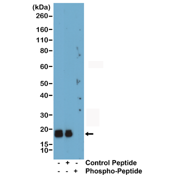
Figure 1. Western blot analysis of Myosin Light Chain 2/MLC-2V/MYL2 using anti-Myosin Light Chain 2/MLC-2V/MYL2 antibody (A03215-3). Electrophoresis was performed on a 5-20% SDS-PAGE gel at 70V (Stacking gel) / 90V (Resolving gel) for 2-3 hours. The sample well of each lane was loaded with 30 ug of sample under reducing conditions. Lane 1: human U20S whole cell lysates, Lane 2: monkey heart tissue lysates, Lane 3: rat heart tissue lysates, Lane 4: rat skeletal muscle tissue lysates, Lane 5: mouse heart tissue lysates, Lane 6: mouse skeletal muscle lysates. After electrophoresis, proteins were transferred to a nitrocellulose membrane at 150 mA for 50-90 minutes. Blocked the membrane with 5% non-fat milk/TBS for 1.5 hour at RT. The membrane was incubated with rabbit anti-Myosin Light Chain 2/MLC-2V/MYL2 antigen affinity purified polyclonal antibody (Catalog # A03215-3) at 0.5 microg/mL overnight at 4°C, then washed with TBS-0.1%Tween 3 times with 5 minutes each and probed with a goat anti-rabbit IgG-HRP secondary antibody at a dilution of 1:5000 for 1.5 hour at RT. The signal is developed using an Enhanced Chemiluminescent detection (ECL) kit (Catalog # EK1002) with Tanon 5200 system. A specific band was detected for Myosin Light Chain 2/MLC-2V/MYL2 at approximately 19 kDa. The expected band size for Myosin Light Chain 2/MLC-2V/MYL2 is at 19 kDa.
Figure 1. Western blot analysis of Myosin Light Chain 2/MLC-2V/MYL2 using anti-Myosin Light Chain 2/MLC-2V/MYL2 antibody (A03215-3). Electrophoresis was performed on a 5-20% SDS-PAGE gel at 70V (Stacking gel) / 90V (Resolving gel) for 2-3 hours. The sample well of each lane was loaded with 30 ug of sample under reducing conditions. Lane 1: human U20S whole cell lysates, Lane 2: monkey heart tissue lysates, Lane 3: rat heart tissue lysates, Lane 4: rat skeletal muscle tissue lysates, Lane 5: mouse heart tissue lysates, Lane 6: mouse skeletal muscle lysates. After electrophoresis, proteins were transferred to a nitrocellulose membrane at 150 mA for 50-90 minutes. Blocked the membrane with 5% non-fat milk/TBS for 1.5 hour at RT. The membrane was incubated with rabbit anti-Myosin Light Chain 2/MLC-2V/MYL2 antigen affinity purified polyclonal antibody (Catalog # A03215-3) at 0.5 microg/mL overnight at 4°C, then washed with TBS-0.1%Tween 3 times with 5 minutes each and probed with a goat anti-rabbit IgG-HRP secondary antibody at a dilution of 1:5000 for 1.5 hour at RT. The signal is developed using an Enhanced Chemiluminescent detection (ECL) kit (Catalog # EK1002) with Tanon 5200 system. A specific band was detected for Myosin Light Chain 2/MLC-2V/MYL2 at approximately 19 kDa. The expected band size for Myosin Light Chain 2/MLC-2V/MYL2 is at 19 kDa.
Anti-Myosin Light Chain 2/MLC-2V/MYL2 Antibody Picoband(r)
A03215-3-CARRIER-FREE
ApplicationsWestern Blot
Product group Antibodies
ReactivityHuman, Monkey, Mouse, Rat
TargetMYL2
Overview
- SupplierBoster Bio
- Product NameAnti-Myosin Light Chain 2/MLC-2V/MYL2 Antibody Picoband(r)
- Delivery Days Customer9
- ApplicationsWestern Blot
- CertificationResearch Use Only
- ClonalityPolyclonal
- Concentration500 ug/ml
- Gene ID4633
- Target nameMYL2
- Target descriptionmyosin light chain 2
- Target synonymsCMH10, MFM12, MLC-2, MLC-2s/v, MLC-2v, MLC2, myosin regulatory light chain 2, ventricular/cardiac muscle isoform, RLC of myosin, S15D myosin regulatory light chain 2, T160D myosin regulatory light chain 2, cardiac myosin light chain 2, cardiac ventricular myosin light chain 2, myosin, light chain 2, regulatory, cardiac, slow, myosin, light polypeptide 2, regulatory, cardiac, slow, regulatory light chain of myosin, slow cardiac myosin regulatory light chain 2, ventricular myosin light chain 2
- HostRabbit
- IsotypeIgG
- Protein IDP10916
- Protein NameMyosin regulatory light chain 2, ventricular/cardiac muscle isoform
- Scientific DescriptionBoster Bio Anti-Myosin Light Chain 2/MLC-2V/MYL2 Antibody Picoband® catalog # A03215-3. Tested in WB applications. This antibody reacts with Human, Monkey, Mouse, Rat. The brand Picoband indicates this is a premium antibody that guarantees superior quality, high affinity, and strong signals with minimal background in Western blot applications. Only our best-performing antibodies are designated as Picoband, ensuring unmatched performance.
- ReactivityHuman, Monkey, Mouse, Rat
- Storage Instruction-20°C,2°C to 8°C
- UNSPSC12352203
Related products
Product group Antibodies
Anti-MYL2 AntibodyA39982
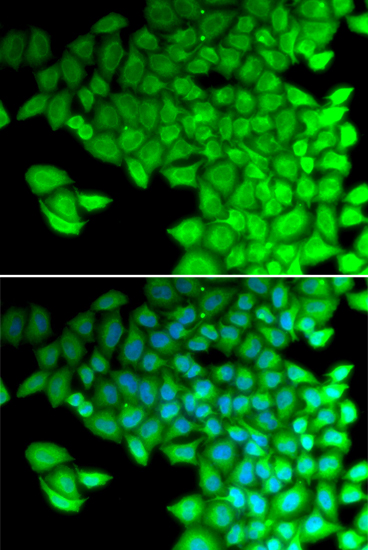
ApplicationsImmunoFluorescence, Western Blot
ReactivityHuman, Mouse, Rat
- SizePrice
Product group Antibodies
Anti-MYL2 Antibody144-60597
ApplicationsWestern Blot
ReactivityHuman, Mouse, Rat
TargetMYL2
- SizePrice
Product group Antibodies
ApplicationsWestern Blot
ReactivityHuman, Mouse, Rat
TargetMYL2
- SizePrice
Product group Antibodies
MYL2 AntibodyCSB-PA10059A0RB
ApplicationsImmunoFluorescence, ELISA, ImmunoHistoChemistry
ReactivityHuman
TargetMYL2
- SizePrice
Product group Antibodies
ApplicationsImmunoPrecipitation, Western Blot, ImmunoCytoChemistry, ImmunoHistoChemistry
ReactivityMouse, Porcine, Rat
TargetMYL2
- SizePrice
Product group Antibodies

MYL2 AntibodyLS-C400902
ApplicationsWestern Blot, ELISA
ReactivityHuman, Mouse, Rat
TargetMYL2
- SizePrice
Product group Antibodies
ApplicationsWestern Blot, ImmunoHistoChemistry, ImmunoHistoChemistry Paraffin
ReactivityHuman, Mouse, Rat
TargetMYL2
- SizePrice

Product group Antibodies
Anti-MYL2 AntibodyHPA019763
ApplicationsWestern Blot, ImmunoHistoChemistry
ReactivityHuman
TargetMYL2
- SizePrice
Product group Antibodies
ApplicationsWestern Blot
ReactivityHuman, Mouse, Rat
TargetMYL2
- SizePrice