

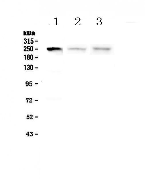
Figure 1. Western blot analysis of MYLK using anti-MYLK antibody (A01697-2). Electrophoresis was performed on a 5-20% SDS-PAGE gel at 70V (Stacking gel) / 90V (Resolving gel) for 2-3 hours. The sample well of each lane was loaded with 50ug of sample under reducing conditions. Lane 1: human SGC-7901 whole cell lysates, Lane 2: rat spleen tissue lysates, Lane 3: mouse spleen tissue lysates. After Electrophoresis, proteins were transferred to a Nitrocellulose membrane at 150mA for 50-90 minutes. Blocked the membrane with 5% Non-fat Milk/ TBS for 1.5 hour at RT. The membrane was incubated with rabbit anti-MYLK antigen affinity purified polyclonal antibody (Catalog # A01697-2) at 0.5 microg/mL overnight at 4°C, then washed with TBS-0.1%Tween 3 times with 5 minutes each and probed with a goat anti-rabbit IgG-HRP secondary antibody at a dilution of 1:10000 for 1.5 hour at RT. The signal is developed using an Enhanced Chemiluminescent detection (ECL) kit (Catalog # EK1002) with Tanon 5200 system. A specific band was detected for MYLK at approximately 250KD. The expected band size for MYLK is at 211KD.
. MYLK was detected in paraffin-embedded section of mouse lung tissue . Heat mediated antigen retrieval was performed in citrate buffer (pH6, epitope retrieval solution) for 20 mins. The tissue section was blocked with 10% goat serum. The tissue section was then incubated with 1microg/ml rabbit anti-MYLK Antibody (A01697-2) overnight at 4°C. Biotinylated goat anti-rabbit IgG was used as secondary antibody and incubated for 30 minutes at 37°C. The tissue section was developed using Strepavidin-Biotin-Complex (SABC)(Catalog # SA1022) with DAB as the chromogen.")
. MYLK was detected in paraffin-embedded section of mouse small intestine tissue. Heat mediated antigen retrieval was performed in citrate buffer (pH6, epitope retrieval solution) for 20 mins. The tissue section was blocked with 10% goat serum. The tissue section was then incubated with 1microg/ml rabbit anti-MYLK Antibody (A01697-2) overnight at 4°C. Biotinylated goat anti-rabbit IgG was used as secondary antibody and incubated for 30 minutes at 37°C. The tissue section was developed using Strepavidin-Biotin-Complex (SABC)(Catalog # SA1022) with DAB as the chromogen.")
. MYLK was detected in paraffin-embedded section of rat small intestine tissue. Heat mediated antigen retrieval was performed in citrate buffer (pH6, epitope retrieval solution) for 20 mins. The tissue section was blocked with 10% goat serum. The tissue section was then incubated with 1microg/ml rabbit anti-MYLK Antibody (A01697-2) overnight at 4°C. Biotinylated goat anti-rabbit IgG was used as secondary antibody and incubated for 30 minutes at 37°C. The tissue section was developed using Strepavidin-Biotin-Complex (SABC)(Catalog # SA1022) with DAB as the chromogen.")
. MYLK was detected in paraffin-embedded section of human placenta tissue . Heat mediated antigen retrieval was performed in citrate buffer (pH6, epitope retrieval solution) for 20 mins. The tissue section was blocked with 10% goat serum. The tissue section was then incubated with 1microg/ml rabbit anti-MYLK Antibody (A01697-2) overnight at 4°C. Biotinylated goat anti-rabbit IgG was used as secondary antibody and incubated for 30 minutes at 37°C. The tissue section was developed using Strepavidin-Biotin-Complex (SABC)(Catalog # SA1022) with DAB as the chromogen.")
. MYLK was detected in paraffin-embedded section of human rectal cancer tissue. Heat mediated antigen retrieval was performed in citrate buffer (pH6, epitope retrieval solution) for 20 mins. The tissue section was blocked with 10% goat serum. The tissue section was then incubated with 1microg/ml rabbit anti-MYLK Antibody (A01697-2) overnight at 4°C. Biotinylated goat anti-rabbit IgG was used as secondary antibody and incubated for 30 minutes at 37°C. The tissue section was developed using Strepavidin-Biotin-Complex (SABC)(Catalog # SA1022) with DAB as the chromogen.")
. MYLK was detected in paraffin-embedded section of rat lung tissue . Heat mediated antigen retrieval was performed in citrate buffer (pH6, epitope retrieval solution) for 20 mins. The tissue section was blocked with 10% goat serum. The tissue section was then incubated with 1microg/ml rabbit anti-MYLK Antibody (A01697-2) overnight at 4°C. Biotinylated goat anti-rabbit IgG was used as secondary antibody and incubated for 30 minutes at 37°C. The tissue section was developed using Strepavidin-Biotin-Complex (SABC)(Catalog # SA1022) with DAB as the chromogen.")
Figure 1. Western blot analysis of MYLK using anti-MYLK antibody (A01697-2). Electrophoresis was performed on a 5-20% SDS-PAGE gel at 70V (Stacking gel) / 90V (Resolving gel) for 2-3 hours. The sample well of each lane was loaded with 50ug of sample under reducing conditions. Lane 1: human SGC-7901 whole cell lysates, Lane 2: rat spleen tissue lysates, Lane 3: mouse spleen tissue lysates. After Electrophoresis, proteins were transferred to a Nitrocellulose membrane at 150mA for 50-90 minutes. Blocked the membrane with 5% Non-fat Milk/ TBS for 1.5 hour at RT. The membrane was incubated with rabbit anti-MYLK antigen affinity purified polyclonal antibody (Catalog # A01697-2) at 0.5 microg/mL overnight at 4°C, then washed with TBS-0.1%Tween 3 times with 5 minutes each and probed with a goat anti-rabbit IgG-HRP secondary antibody at a dilution of 1:10000 for 1.5 hour at RT. The signal is developed using an Enhanced Chemiluminescent detection (ECL) kit (Catalog # EK1002) with Tanon 5200 system. A specific band was detected for MYLK at approximately 250KD. The expected band size for MYLK is at 211KD.
Anti-Myosin light chain kinase/MYLK Antibody Picoband(r)
A01697-2-CARRIER-FREE
ApplicationsWestern Blot, ELISA, ImmunoHistoChemistry
Product group Antibodies
ReactivityHuman, Mouse, Rat
TargetMYLK
Overview
- SupplierBoster Bio
- Product NameAnti-Myosin light chain kinase/MYLK Antibody Picoband(r)
- Delivery Days Customer9
- ApplicationsWestern Blot, ELISA, ImmunoHistoChemistry
- CertificationResearch Use Only
- ClonalityPolyclonal
- Concentration500 ug/ml
- Gene ID4638
- Target nameMYLK
- Target descriptionmyosin light chain kinase
- Target synonymsAAT7, KRP, MLCK, MLCK1, MLCK108, MLCK210, MMIHS, MMIHS1, MSTP083, MYLK-L, MYLK1, smMLCK, myosin light chain kinase, smooth muscle, kinase-related protein, myosin, light polypeptide kinase, smooth muscle myosin light chain kinase, telokin
- HostRabbit
- IsotypeIgG
- Protein IDQ15746
- Protein NameMyosin light chain kinase, smooth muscle
- Scientific DescriptionBoster Bio Anti-Myosin light chain kinase/MYLK Antibody Picoband® catalog # A01697-2. Tested in ELISA, IHC, WB applications. This antibody reacts with Human, Mouse, Rat. The brand Picoband indicates this is a premium antibody that guarantees superior quality, high affinity, and strong signals with minimal background in Western blot applications. Only our best-performing antibodies are designated as Picoband, ensuring unmatched performance.
- ReactivityHuman, Mouse, Rat
- Storage Instruction-20°C,2°C to 8°C
- UNSPSC12352203
Related products
Product group Antibodies
Anti-MYLK AntibodyA98020
ApplicationsWestern Blot, ELISA
ReactivityHuman, Mouse, Rat
- SizePrice
Product group Antibodies

MYLK AntibodyLS-C682218
ApplicationsImmunoFluorescence, ELISA, ImmunoHistoChemistry, ImmunoHistoChemistry Paraffin
ReactivityHuman
TargetMYLK
- SizePrice
Product group Antibodies

Goat anti-MYLKEB07865
ApplicationsWestern Blot, ELISA
ReactivityHuman, Mouse
TargetMYLK
- SizePrice
Product group Antibodies
Mylk Polyclonal AntibodyCAC11090
ApplicationsImmunoFluorescence, ELISA, ImmunoHistoChemistry
TargetMYLK
- SizePrice
Product group Antibodies
Phospho-MYLK (Y464) AntibodyCSB-PA020005
ApplicationsELISA, ImmunoHistoChemistry
ReactivityHuman
TargetMYLK
- SizePrice
Product group Antibodies
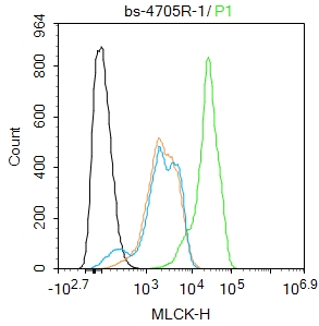
MLCK Polyclonal AntibodyBS-4705R
ApplicationsImmunoFluorescence, ELISA, ImmunoCytoChemistry, ImmunoHistoChemistry, ImmunoHistoChemistry Frozen, ImmunoHistoChemistry Paraffin
ReactivityBovine, Canine, Chicken, Equine, Guinea Pig, Human, Mouse, Porcine, Rat, Sheep
TargetMYLK
- SizePrice
Product group Antibodies
MLCK (phospho Tyr464) antibodyGTX03352
ApplicationsWestern Blot, ELISA
ReactivityHuman
TargetMYLK
- SizePrice

Product group Antibodies
Anti-MYLK AntibodyHPA031677
ApplicationsWestern Blot, ImmunoHistoChemistry
ReactivityHuman
TargetMYLK
- SizePrice
Product group Antibodies
TargetMYLK
- SizePrice
Product group Antibodies
ApplicationsWestern Blot, ELISA
ReactivityHuman
TargetMYLK
- SizePrice